
Image: Colonic crypts within four tissue sections

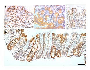
Description: Colonic crypts (intestinal glands) within four tissue sections. The cells have been stained by immunohistochemistry to show a brown-orange color if the cells produce the mitochondrial protein Cytochrome c oxidase subunit I (CCOI), and the nuclei of the cells (located at the outer edges of the cells lining the walls of the crypts) are stained blue-gray with haematoxylin. Panels A, B were cut across the long axes of the crypts and panels C, D were cut parallel to the long axes of the crypts. In panel A the bar shows 100 µm and allows an estimate of the frequency of crypts in the colonic epithelium. Panel B includes three crypts in cross-section, each with one segment deficient for CCOI expression and at least one crypt, on the right side, undergoing fission into two crypts. Panel C shows, on the left side, a crypt fissioning into two crypts. Panel D shows typical small clusters of two and three CCOI deficient crypts (the bar shows 50 µm). The images were made from original photomicrographs, but panels A, B and D were also included in an article[1] and illustrations were published with Creative Commons Attribution-Noncommercial License allowing re-use. ↑ Template:Vcite2 journal
Title: Colonic crypts within four tissue sections
Credit: Own work
Author: Chaya5260
Usage Terms: Creative Commons Attribution-Share Alike 4.0
License: CC BY-SA 4.0
License Link: http://creativecommons.org/licenses/by-sa/4.0
Attribution Required?: Yes
Image usage
The following page links to this image:
