
Image: 3D CT of thorax, annotated
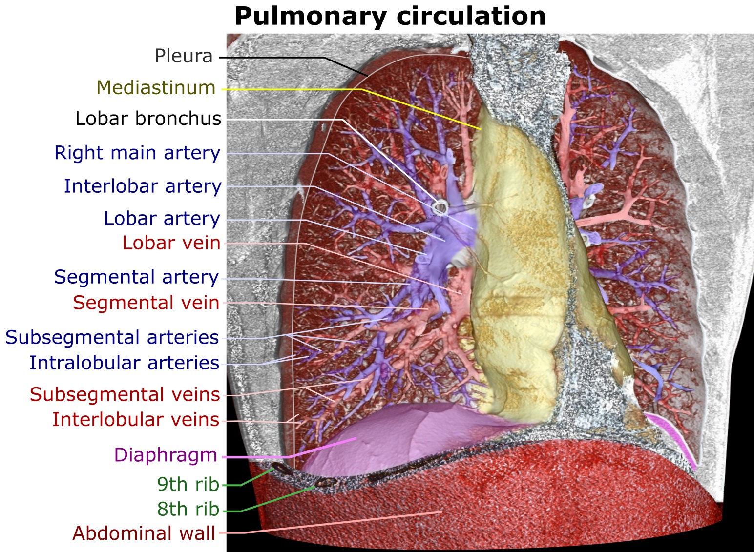
Description: edit Artificially colored 3D rendering of a high resolution computed tomography of a normal thorax of a 37 year old man who presented with unspecific breathing problems, published with written informed consent. The anterior thoracic wall, the airways and the pulmonary vessels anterior to the root of the lung have been digitally removed in order to visualize the different levels of the pulmonary circulation.
Title: 3D CT of thorax, annotated
Credit: Own work edit References: Main pulmonary artery, segmental and subsegmental pulmonary arteries: Pulmonary Artery Anatomy. University of Virginia School of Medicine (2013). Retrieved on 2017-06-24. Right and left main pulmonary artery: Pulmonary Vasculature. University of Virginia School of Medicine (2013). Retrieved on 2017-06-24. Intralobular arteries: (2008). "Imaging of pulmonary emphysema: a pictorial review". Int J Chron Obstruct Pulmon Dis 3 (2): 193–204. PMID 18686729. PMC: 2629965. Interlobular veins: Page 58 in: David H. Trapnell (2016) Principles of X-Ray Diagnosis, Butterworth-Heinemann ISBN: 9781483195384.
Author: Mikael Häggström
Usage Terms: Creative Commons Zero, Public Domain Dedication
License: CC0
License Link: http://creativecommons.org/publicdomain/zero/1.0/deed.en
Attribution Required?: No
Image usage
The following 4 pages link to this image:
