
Image: Atypical leiomyoma intermed mag
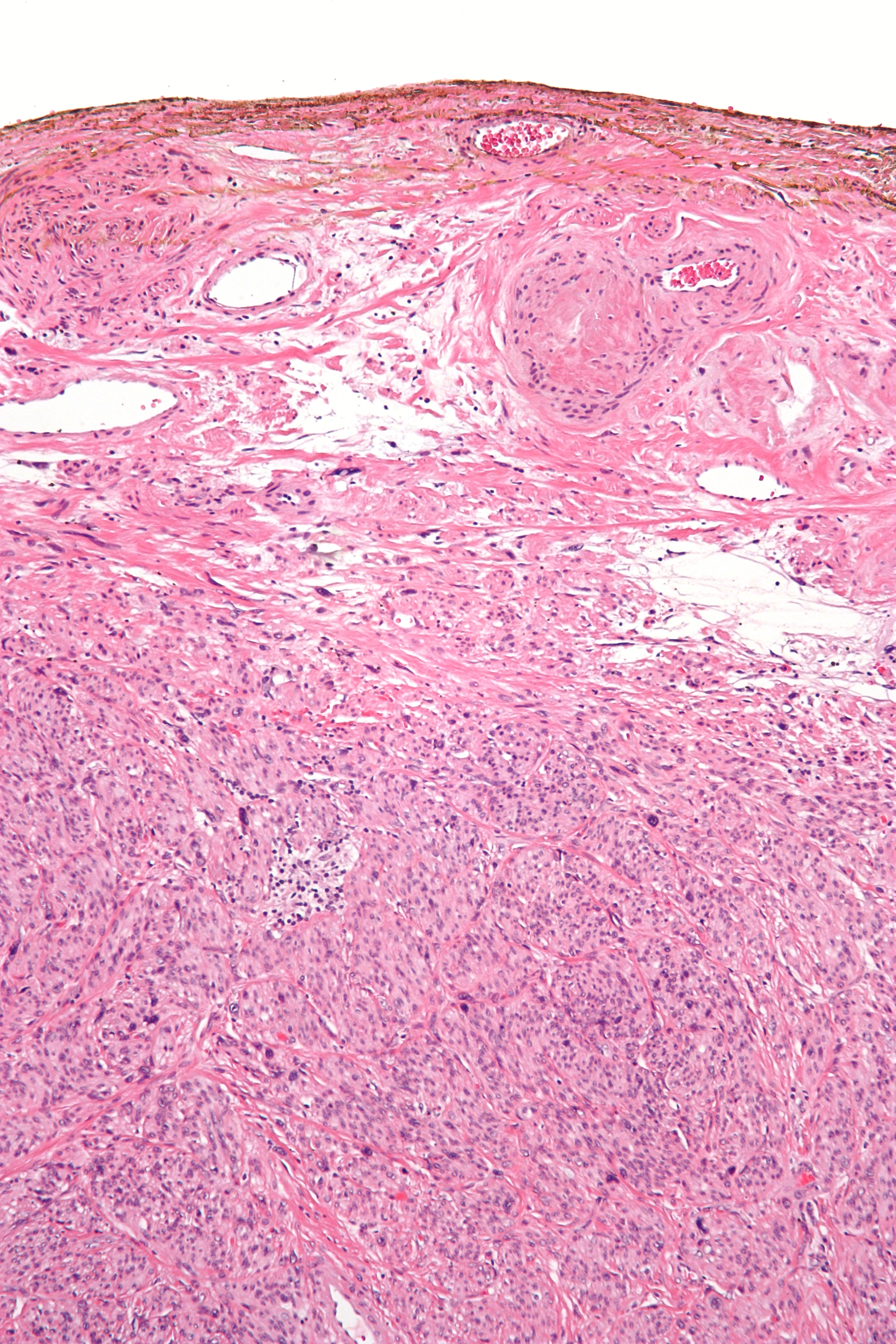
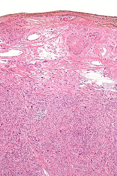
Description: Intermediate magnification micrograph of an atypical leiomyoma, also known as an atypical fibroid and symplastic leiomyoma. Surgical excision specimen. H&E stain. The resection margin was painted with silver nitrate. The image shows a mixed cell population consisting of epithelioid cells and spindle cells with marked nuclear atypia. Mitotic activity and necrosis is absent. The presence of mitotic activity or necrosis in addition to nuclear atypia would make the lesion a leiomyosarcoma. See also Image:Atypical leiomyoma low mag.jpg Image:Atypical leiomyoma high mag.jpg
Title: Atypical leiomyoma intermed mag
Credit: Own work
Author: Nephron
Usage Terms: Creative Commons Attribution-Share Alike 3.0
License: CC BY-SA 3.0
License Link: http://creativecommons.org/licenses/by-sa/3.0
Attribution Required?: Yes
Image usage
The following page links to this image:
