
Image: Blood clot in scanning electron microscopy
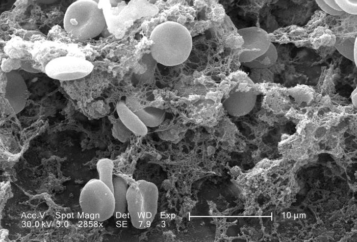
Description: This scanning electron micrograph (SEM) depicted a number of red blood cells found enmeshed in a fibrinous matrix on the luminal surface of an indwelling vascular catheter; Magnified 2858x. Note the biconcave cytomorphologic shape of each erythrocyte, which increases the surface area of these hemoglobin-filled cells, thereby, promoting a greater degree of gas exchange, which is their primary function in an in vivo setting. In their adult phase, these cells possess no nucleus. What appears to be irregularly-shaped chunks of debris, are actually fibrin clumps, which when inside the living organism, functions as a key component in the process of blood clot formation, acting to entrap the red blood cells in a mesh-like latticework of proteinaceous strands, thereby, stabilizing and strengthening the clot, in much the same way as rebar acts to strengthen, and reinforce cement.
Title: Blood clot in scanning electron microscopy
Credit: http://phil.cdc.gov/phil/details.asp
Author: Janice Carr
Usage Terms: Public domain
License: Public domain
Attribution Required?: No
Image usage
The following page links to this image:
