
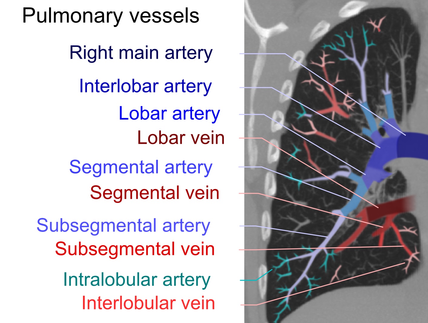
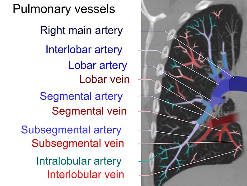
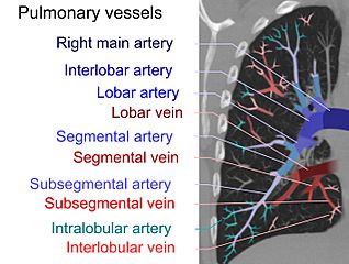
Image: Computed tomograph of pulmonary vessels
Description: Artificially colored high resolution computed tomography of a normal lung, using a 1 cm thick maximum intensity projection in order to visualize the different levels of pulmonary arteries and veins.
Title: Computed tomograph of pulmonary vessels
Credit: Own work edit References: Main pulmonary artery, segmental and subsegmental pulmonary arteries: Pulmonary Artery Anatomy. University of Virginia School of Medicine (2013). Retrieved on 2017-06-24. Right and left main pulmonary artery: Pulmonary Vasculature. University of Virginia School of Medicine (2013). Retrieved on 2017-06-24. Intralobular arteries: (2008). "Imaging of pulmonary emphysema: a pictorial review". Int J Chron Obstruct Pulmon Dis 3 (2): 193–204. PMID 18686729. PMC: 2629965. Interlobular veins: Page 58 in: David H. Trapnell (2016) Principles of X-Ray Diagnosis, Butterworth-Heinemann ISBN: 9781483195384.
Author: Mikael Häggström
Usage Terms: Creative Commons Zero, Public Domain Dedication
License: CC0
License Link: http://creativecommons.org/publicdomain/zero/1.0/deed.en
Attribution Required?: No
Image usage
The following page links to this image:
