
Image: Helix electron density myoglobin 2nrl 17-32
Size of this preview: 341 × 599 pixels. Other resolutions: 136 × 240 pixels | 740 × 1,300 pixels.
Original image (740 × 1,300 pixels, file size: 356 KB, MIME type: image/jpeg)
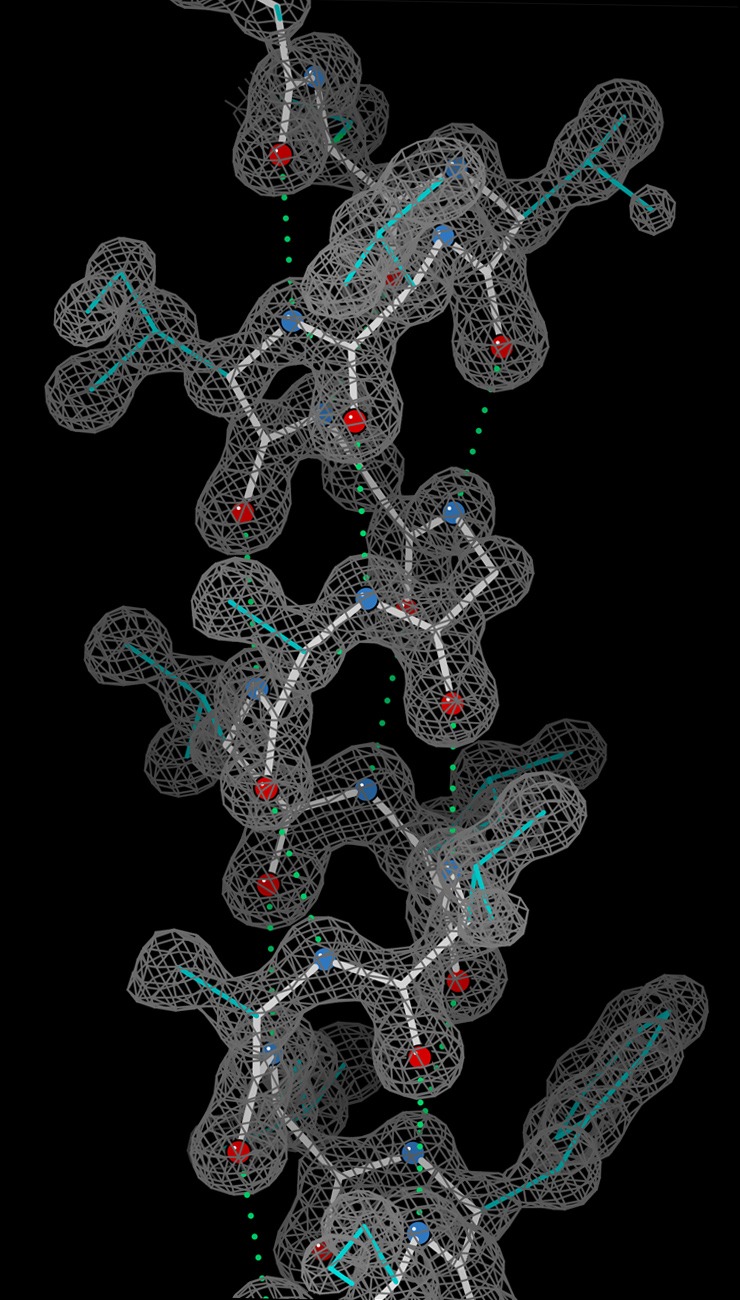
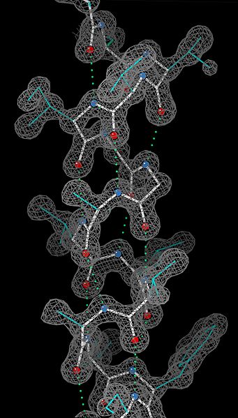
Description: An alpha-helix, with stick-figures for the model shown within electron density for the crystal structure at ultra-high-resolution (0.91Å). The density contours are in gray, the helix backbone in white, sidechains in cyan, O atoms in red, N atoms in blue, and hydrogen bonds as green dotted lines. From PDB file 2NRL, residues 17-32.
Title: Helix electron density myoglobin 2nrl 17-32
Credit: Own work
Author: Dcrjsr
Usage Terms: Creative Commons Attribution 3.0
License: CC BY 3.0
License Link: http://creativecommons.org/licenses/by/3.0
Attribution Required?: Yes
Image usage
The following 3 pages link to this image:

All content from Kiddle encyclopedia articles (including the article images and facts) can be freely used under Attribution-ShareAlike license, unless stated otherwise.