
Image: MHC Binding Diagram
Size of this preview: 800 × 545 pixels. Other resolutions: 320 × 218 pixels | 1,098 × 748 pixels.
Original image (1,098 × 748 pixels, file size: 66 KB, MIME type: image/png)
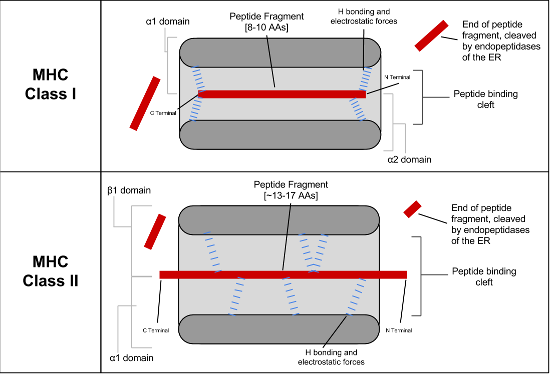
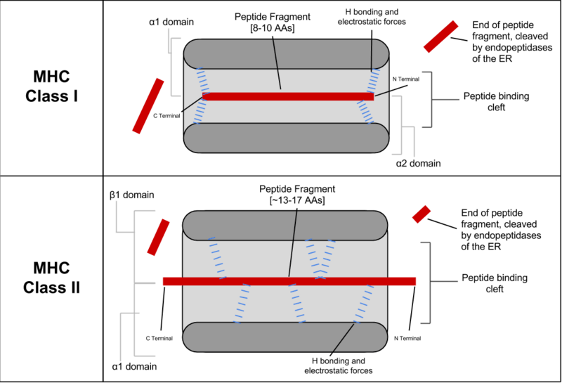
Description: Peptide binding for Class I and Class II MHC molecules, showing the binding of peptides between the alpha-helix walls, upon a beta-sheet base. The difference in binding positions is shown. Class I primarily makes contact with backbone residues at the Carboxy and amino terminal regions, while Class II primarily makes contacts along the length of the residue backbone. The precise location of binding residues is determined by the MHC allele.
Title: MHC Binding Diagram
Credit: Own work
Author: Connor Sampson
Usage Terms: Creative Commons Attribution-Share Alike 4.0
License: CC BY-SA 4.0
License Link: http://creativecommons.org/licenses/by-sa/4.0
Attribution Required?: Yes
Image usage
The following page links to this image:

All content from Kiddle encyclopedia articles (including the article images and facts) can be freely used under Attribution-ShareAlike license, unless stated otherwise.