
Image: Parasite130103-fig1 Protopolystoma xenopodis (Monogenea, Polystomatidae) egg

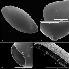
Description: Scanning electron micrographs of Protopolystoma xenopodis egg features. (A) Fully embryonated egg, operculum visible (← Op). (B) Operculum becoming visible as the egg develops (← Op). (C) A residual structure on the non-opercular side of the egg. (D) An empty egg shell after the oncomiracidium has left. (E) Egg shell indicating the thickness of an individual parasite egg shell at the opercular opening.
Title: Parasite130103-fig1 Protopolystoma xenopodis (Monogenea, Polystomatidae) egg
Credit: Theunissen, M., Tiedt, L. & Du Preez, L. H. 2014: The morphology and attachment of Protopolystoma xenopodis (Monogenea: Polystomatidae) infecting the African clawed frog Xenopus laevis. Parasite, 21, 20. doi:10.1051/parasite/2014020
Author: Maxine Theunissen, Louwrens Tiedt and Louis H. Du Preez
Usage Terms: Creative Commons Attribution 2.0
License: CC BY 2.0
License Link: http://creativecommons.org/licenses/by/2.0
Attribution Required?: Yes
Image usage
There are no pages that link to this image.
