
Image: Pterosaur respiratory system
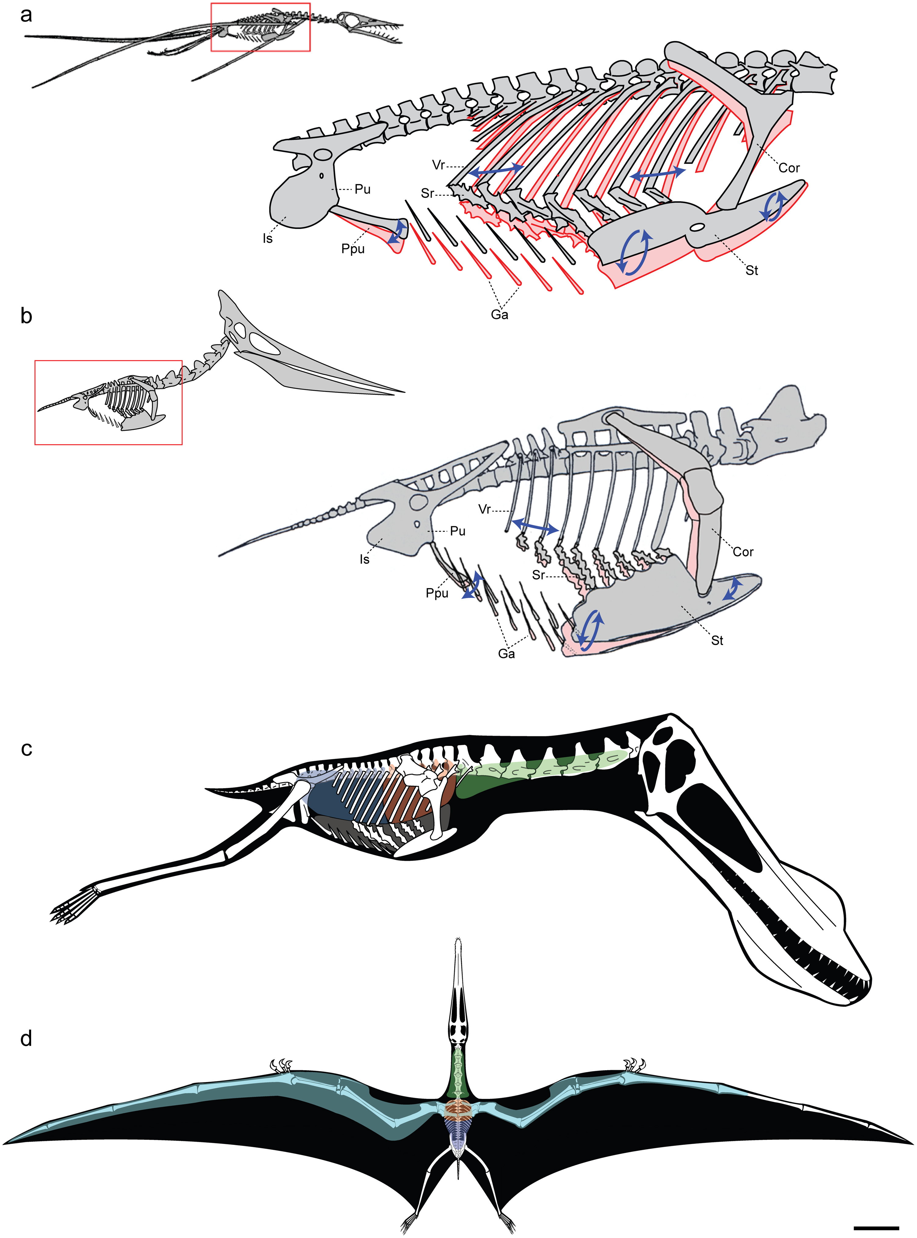
Description: Models of ventilatory kinematics and the pulmonary air sac system of pterosaurs. a, Model of ventilatory kinematics in Rhamphorhynchus. Thoracic movement induced by the ventral intercostal musculature results in forward and outward displacement of the distal vertebral and proximal sternal ribs, and ventral displacement of the sternum, upon inspiration (blue arrows and pink outline). In addition, ventral expansion of the abdomen is induced through caudoventral rotation of the prepubis. Ranges of skeletal movement were modelled after those observed in vivo in the avian thorax and the crocodylian pelvis [26], [27]. Rhamphorhynchus modified from Wellnhofer [48]. b, Model of ventilatory kinematics in Pteranodon wherein the fused anterior vertebral ribs and articulation of the scapulocoracoid with the supraneural plate and anterior sternum limit movement of the anterior sternum, which cannot undergo elliptical rotation. However, the posterior vertebral ribs, sternal ribs, sternum, and prepubis are still capable of anterodorsal-posteroventral excursions facilitating volumetric increases and decreases of the thorax during inspiration-expiration. Pteranodon modified from Bennett [29]. c, d, reconstruction of pulmonary air sac system in the Lower Cretaceous ornithocheirid Anhanguera santanae (AMNH 22555). c, Lateral view showing the inferred position of the lungs (orange), cervical (green) and abdominal air sacs (blue), as predicted on the basis of postcranial skeletal pneumaticity. Thoracic air sacs (shown in grey) are also likely to have been present, but generally do not leave a distinct osteological trace. Humerus and more distal forelimb not shown. d, Dorsal view illustrating the inferred position of subcutaneous diverticular networks (light blue) distally along the wing. The right side depicts a conservative estimate for the size of the airsac network, limiting it to the pre-axial margin of the wing based solely on the presence of pneumatic foramina in closely positioned wing bones. The left side depicts the likely maximal size of an inferred diverticular network, accounting for its inclusion between the dorsal and ventral layers of the wing membrane. Scale = 10 cm. Skeletal reconstruction in c, d modified from Wellnhofer [49]. Abbreviations: as in figure 2, and: Cor: coracoid portion of scapulocoracoid, Ga: gastralia.
Title: Pterosaur respiratory system
Credit: http://www.plosone.org/article/info%3Adoi%2F10.1371%2Fjournal.pone.0004497;jsessionid=A57F0FDB595AC49992E2B5A390FA104C
Author: Leon P. A. M. Claessens, Patrick M. O'Connor, David M. Unwin
Usage Terms: Creative Commons Attribution 2.5
License: CC BY 2.5
License Link: https://creativecommons.org/licenses/by/2.5
Attribution Required?: Yes
Image usage
The following image is a duplicate of this image (more details):
The following page links to this image:
