
Image: Spindle centriole - embryonic brain mouse - TEM

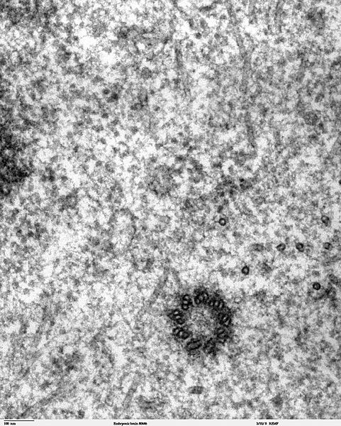
Description: Transmission electron microscope image of a thin section cut through the developing brain tissue (telencephalic hemisphere) of an 11.5 day mouse embryo. This high magnification image of "Embryonic brain 80445" show a spindle centriole and some spindle microtubules visible in the cytoplasm of a mitotic cell at the luminal surface of the telencephalon. JEOL 100CX TEM References: Marin-Padilla, M. (1985) "Early Vascularization of the Embryonic Cerebral Cortex: Golgi and Electron Microscope Studies", J. Comparative Neurology, 241:237-249 Marin-Padilla, M. and M. Amievo (1989) "Early Neurogenesis of the Mouse Olfactory Nerve: Golgi and Electron Microscope Studies", J. Comparative Neurology, 288:339-352
Title: Spindle centriole - embryonic brain mouse - TEM
Credit: http://remf.dartmouth.edu/imagesindex.html http://remf.dartmouth.edu/images/mammalianBrainTEM/source/15.html
Author: Louisa Howard, Miguel Marin-Padilla
Permission: PD
Usage Terms: Public domain
License: Public domain
Attribution Required?: No
Image usage
The following page links to this image:
