
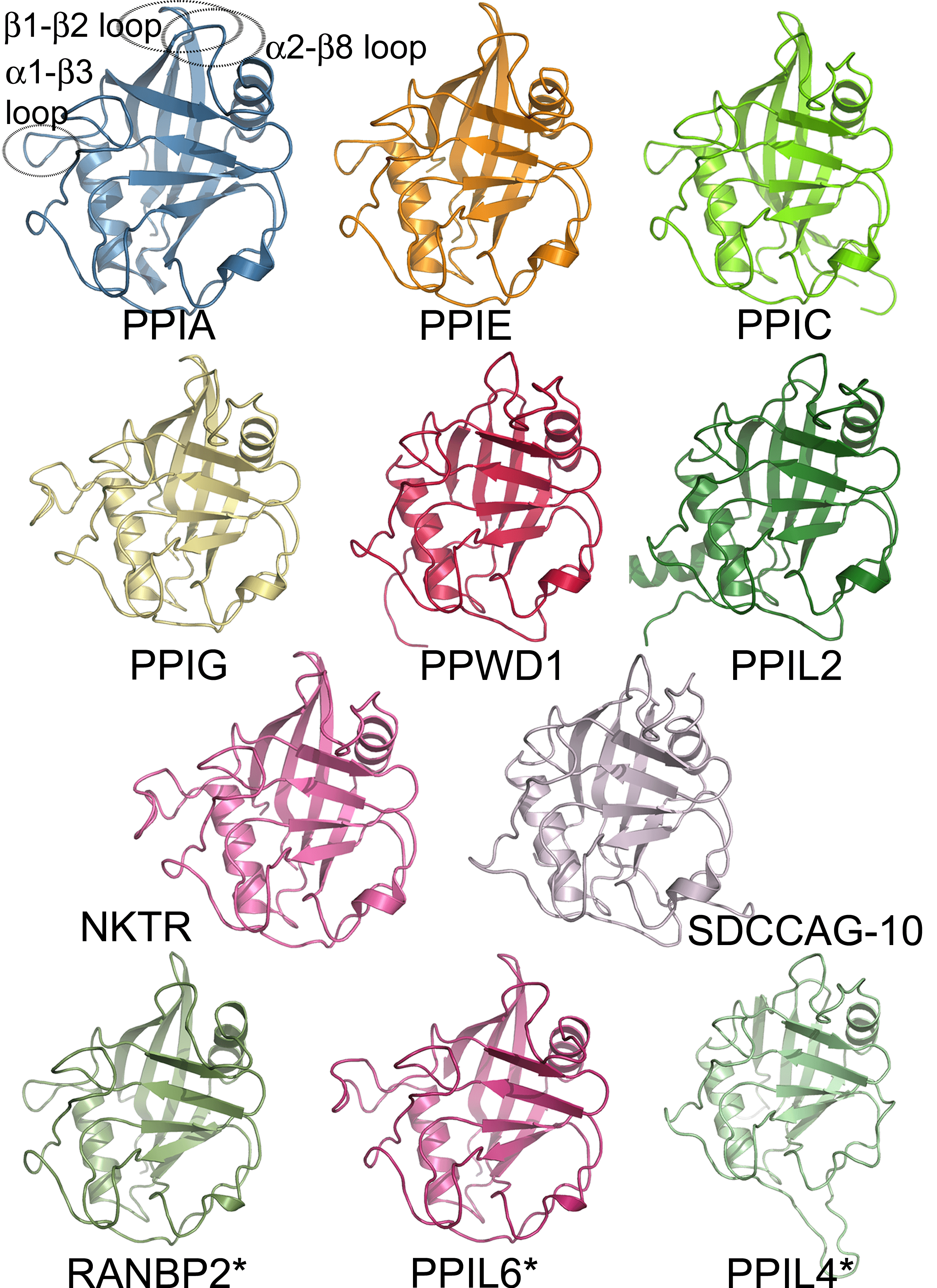
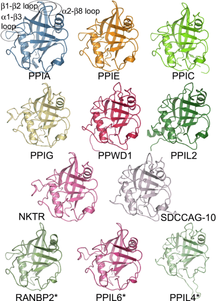
Image: Structural coverage of the human cyclophilin family
Description: Cartoon representation of the novel experimental and modeled structures of human cyclophilins associated with Davis et al., PLoS Biol 8(7): e1000439, 2010. Only the isomerase domain is shown. The previously determined structure of PPIA is shown as a reference point, and loop regions discussed in the text are outlined with dotted ovals and labeled. The structures of RanBP2, PPIL6, and PPIL4 are marked with an asterisk, as they are derived from homology modeling and do not represent experimentally derived data.
Title: Structural coverage of the human cyclophilin family
Credit: Fig. 2 in Davis et al., PLoS Biol 8(7): e1000439, 2010
Author: Davis et al., PLoS Biol 8(7): e1000439, 2010
Usage Terms: Creative Commons Attribution 2.5
License: CC BY 2.5
License Link: http://creativecommons.org/licenses/by/2.5
Attribution Required?: Yes
Image usage
The following page links to this image:
