
Image: Tasmaniosaurus misc. postcranial
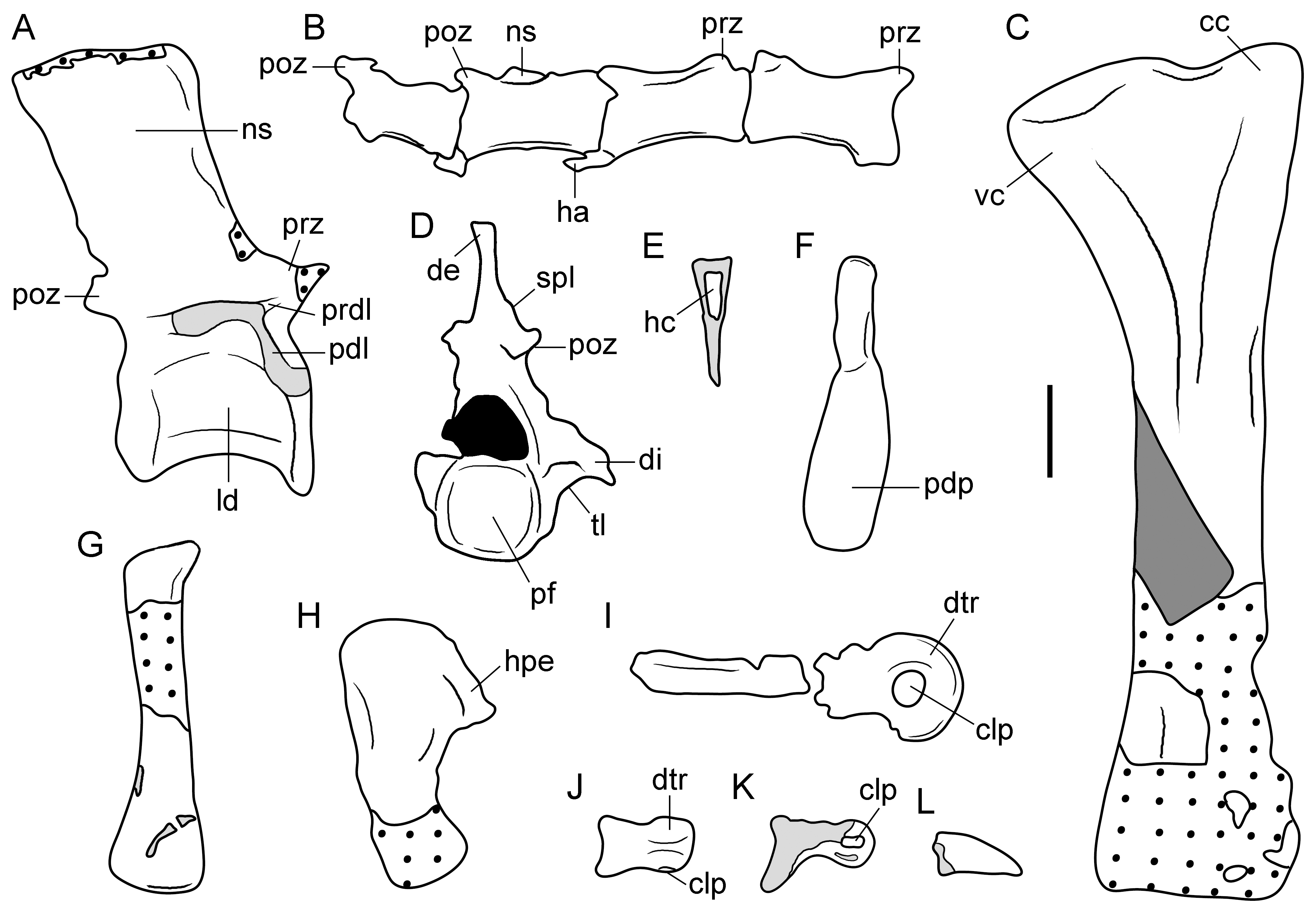
Description: Figure 10. Line drawings of selected postcranial bones of type specimen (UTGD 54655) of Tasmaniosaurus triassicus. A, anterior or middle dorsal vertebra and B, middle caudal vertebrae in right lateral views; C, tibia in lateral or medial view; D, cervico-dorsal vertebra in posterior view; E, proximal half of haemal arch in crosss-section; F, anterior or middle haemal arch in right lateral view; G, probable metatarsal II and H, metatarsal V in dorsal or ventral views; I, proximal pedal phalanx in side view; J, pedal phalanx in ventral view; K, pedal phalanx in side view; and L, ungueal pedal phalanx in side view. Areas with dotted lines are bone impressions, light grey areas are damaged bone, and neural canal in black. Abbreviations: cc, cnemial crest; clp, collateral pit; de, possibly artificial distal transverse expansion; di, diapophysis; dtr, distal trochlea; ha, haemal arch; hc, haemal canal; hpe, hook-shaped proximal end; ld, lateral depression in the centrum; ns, neural spine; pdl, paradiapophyseal lamina; pdp, plate-like distal end; pf, posterior articular surface; poz, postzygapophysis; prz, prezygapophysis; prdl, prezygodiapophyseal lamina; spl, spinopostzygapophyseal lamina; tl, thick lamina; vc, ventral condyle. Scale bar equals 1 cm.
Title: Tasmaniosaurus misc. postcranial
Credit: The Osteology of the Basal Archosauromorph Tasmaniosaurus triassicus from the Lower Triassic of Tasmania, Australia. PLoS ONE 9(1): e86864. https://doi.org/10.1371/journal.pone.0086864
Author: Martin D. Ezcurra (2014)
Usage Terms: Creative Commons Attribution 4.0
License: CC BY 4.0
License Link: https://creativecommons.org/licenses/by/4.0
Attribution Required?: Yes
Image usage
The following page links to this image:
