
Eye facts for kids


The eye is a round organ for sensing light so animals can see. About 97 percent of animals have eyes. Image-resolving eyes are present in cnidaria, molluscs, vertebtates, annelids and arthropods.
In mammals, two kinds of cells, rods and cones, allow sight by sending signals through the optic nerve to the brain.
Some animals can see light that humans cannot see. They can see ultraviolet or infrared light.
The lens on the front part of the eye acts like a camera lens. It can be pulled flatter by muscles inside the eye, or allowed to become rounder. As some people get older, they may not be as able to do this perfectly. Many people are born with other small problems or get them later in life, and they may need eyeglasses (or contact lenses) to fix the problem.
Contents
Types of eye
Today, ten different types of eyes are known. Most ways of capturing an image have evolved at least once.
One way to categorize eyes is to look at the number of "chambers". Simple eyes are made of only one concave chamber, perhaps with a lens. Compound eyes have many such chambers with their lenses on a convex surface.
Eyes also can be grouped according to how the photoreceptor is made. Photoreeptors are either cillated, or rhabdomic. and some annelids possess both.
Simple eyes
Pit eyes
Pit eyes are set in a depression in the skin. This reduces the angles at which light can enter. It allows the organism to say where the light is coming from.
Such eyes can be found in about 85% of phyla. They probably came before the development of more complex eyes. Pit eyes are small. They are made of up to about hundred cells, covering about 100 µm. The directionality can be improved by reducing the size of the opening, and by putting a reflective layer behind the receptor cells.
Pinhole eye
.jpg)
The pinhole eye is an advanced form of pit eye. It has several bits, most notably a small aperture and deep pit. Sometimes, the aperture can be changed. It is only found in the Nautilus. Without a lens to focus the image, it produces a blurry image. Consequently, nautiloids can not discriminate between objects with a separation of less than 11°. Shrinking the aperture would produce a sharper image, but let in less light.
Spherical lensed eye
The resolution of pit eyes can be improved a lot by adding a material to make a lens. This will reduce the radius of the blurring, and increase the resolution that can be achieved. The most basic form can still be seen in some gastropods and annelids. These eyes have a lens of one refractive index. It is possible to get a better image with materials that have a high refractive index which decreases towards the edges. This decreases the focal length and allows a sharp image to form on the retina.
This eye creates an image that is sharp enough that motion of the eye can cause significant blurring. To minimize the effect of eye motion while the animal moves, most such eyes have stabilizing eye muscles.
The ocelli of insects have a simple lens, but their focal point always lies behind the retina.They can never form a sharp image. This limits the function of the eye. Ocelli (pit-type eyes of arthropods) blur the image across the whole retina. They are very good at responding to rapid changes in light intensity across the whole visual field — this fast response is accelerated even more by the large nerve bundles which rush the information to the brain. Focusing the image would also cause the sun's image to be focused on a few receptors. These could possibly be damaged by the intense light; shielding the receptors would block out some light and reduce their sensitivity.
This fast response has led to suggestions that the ocelli of insects are used mainly in flight, because they can be used to detect sudden changes in which way is up (because light, especially UV light which is absorbed by vegetation, usually comes from above).
Refractive cornea
The eyes of most land-living vertebrates (as well as those of some spiders, and insect larvae) contain a fluid that has a higher refractive index than the air. That way, the lens does not have to reduce the focal length, because this is done by the fluid. That way, the lens can adjust the focus more easily. That way, a very high resolution can be obtained.
Reflector eyes

Instead of using a lens it is also possible to have cells inside the eye that act like mirrors. The image can then be reflected to focus at a central point. This design also means that someone looking into such an eye will see the same image as the organism which has them.
Many small organisms such as rotifers, copeopods and platyhelminthes use such this design, but their eyes are too small to produce usable images. Some larger organisms, such as scallops, also use reflector eyes. The scallop Pecten has up to 100 millimeter-scale reflector eyes fringing the edge of its shell. It detects moving objects as they pass successive lenses.
Compound eyes
-
Main page: Compound eye

Compound eyes are different from simple eyes. Instead of having one organ that can sense light, they put together many such organs. Some compound eyes have thousands of them. The resulting image is put together in the brain, based on the signals of the many eye units. Each such unit is called ommatidium, several are called ommatidia. The ommatidia are located on a convex surface, each of them points in a slighly different direction. Unlike simple eyes, compound eyes have a very large angle of view. They can detect fast movement, and sometimes the polarization of light.
Compound eyes are common in arthropods, annelids, and some bivalved molluscs
Evolution of the eye
The evolution of eyes started with simplest light-sensitive patches in unicellular organisms. These eye-spots do nothing but detect if the surroundings are light or dark. Most animals have a biochemical 'clock' inside. These simple eye-spots are used to adjust this daily clock, which is called circadian rhythm. Some snails, for example, see no image (picture) at all, but they sense light, which helps them stay out of bright sunlight.
More complex eyes have not lost this function. A special type of cells in the eye senses light for a different purpose than seeing. These cells are called ganglion cells. They are located in the retina. They send their information about light to the brain along a different path (the retinohypothalamic tract). This information adjusts (synchronizes) the animal's circadian rhythm to nature's light/dark cycle of 24 hours. The system also works for some blind people who cannot see light at all.
Eyes that are a little bit better are shaped like cups, which lets the animal know where the light is coming from.
More complex eyes give the full sense of vision, including color, motion, and texture. These eyes have a round shape that makes light rays focus on the back part of the eye, called the retina.
Physiology
Visual acuity

Visual acuity, or resolving power, is "the ability to distinguish fine detail" and is the property of cone cells. It is often measured in cycles per degree (CPD), which measures an angular resolution, or how much an eye can differentiate one object from another in terms of visual angles. Resolution in CPD can be measured by bar charts of different numbers of white/black stripe cycles. For example, if each pattern is 1.75 cm wide and is placed at 1 m distance from the eye, it will subtend an angle of 1 degree, so the number of white/black bar pairs on the pattern will be a measure of the cycles per degree of that pattern. The highest such number that the eye can resolve as stripes, or distinguish from a grey block, is then the measurement of visual acuity of the eye.
For a human eye with excellent acuity, the maximum theoretical resolution is 50 CPD (1.2 arcminute per line pair, or a 0.35 mm line pair, at 1 m). A rat can resolve only about 1 to 2 CPD. A horse has higher acuity through most of the visual field of its eyes than a human has, but does not match the high acuity of the human eye's central fovea region.
Spherical aberration limits the resolution of a 7 mm pupil to about 3 arcminutes per line pair. At a pupil diameter of 3 mm, the spherical aberration is greatly reduced, resulting in an improved resolution of approximately 1.7 arcminutes per line pair. A resolution of 2 arcminutes per line pair, equivalent to a 1 arcminute gap in an optotype, corresponds to 20/20 (normal vision) in humans.
However, in the compound eye, the resolution is related to the size of individual ommatidia and the distance between neighbouring ommatidia. Physically these cannot be reduced in size to achieve the acuity seen with single lensed eyes as in mammals. Compound eyes have a much lower acuity than vertebrate eyes.
Colour perception

"Colour vision is the faculty of the organism to distinguish lights of different spectral qualities." All organisms are restricted to a small range of electromagnetic spectrum; this varies from creature to creature, but is mainly between wavelengths of 400 and 700 nm. This is a rather small section of the electromagnetic spectrum, probably reflecting the submarine evolution of the organ: water blocks out all but two small windows of the EM spectrum, and there has been no evolutionary pressure among land animals to broaden this range.
The most sensitive pigment, rhodopsin, has a peak response at 500 nm. Small changes to the genes coding for this protein can tweak the peak response by a few nm; pigments in the lens can also filter incoming light, changing the peak response. Many organisms are unable to discriminate between colours, seeing instead in shades of grey; colour vision necessitates a range of pigment cells which are primarily sensitive to smaller ranges of the spectrum. In primates, geckos, and other organisms, these take the form of cone cells, from which the more sensitive rod cells evolved. Even if organisms are physically capable of discriminating different colours, this does not necessarily mean that they can perceive the different colours; only with behavioural tests can this be deduced.
Most organisms with colour vision are able to detect ultraviolet light. This high energy light can be damaging to receptor cells. With a few exceptions (snakes, placental mammals), most organisms avoid these effects by having absorbent oil droplets around their cone cells. The alternative, developed by organisms that had lost these oil droplets in the course of evolution, is to make the lens impervious to UV light — this precludes the possibility of any UV light being detected, as it does not even reach the retina.
Rods and cones

The retina contains two major types of light-sensitive photoreceptor cells used for vision: the rods and the cones.
Rods cannot distinguish colours, but are responsible for low-light (scotopic) monochrome (black-and-white) vision; they work well in dim light as they contain a pigment, rhodopsin (visual purple), which is sensitive at low light intensity, but saturates at higher (photopic) intensities. Rods are distributed throughout the retina but there are none at the fovea and none at the blind spot. Rod density is greater in the peripheral retina than in the central retina.
Cones are responsible for colour vision. They require brighter light to function than rods require. In humans, there are three types of cones, maximally sensitive to long-wavelength, medium-wavelength, and short-wavelength light (often referred to as red, green, and blue, respectively, though the sensitivity peaks are not actually at these colours). The colour seen is the combined effect of stimuli to, and responses from, these three types of cone cells. Cones are mostly concentrated in and near the fovea. Only a few are present at the sides of the retina. Objects are seen most sharply in focus when their images fall on the fovea, as when one looks at an object directly. Cone cells and rods are connected through intermediate cells in the retina to nerve fibres of the optic nerve. When rods and cones are stimulated by light, they connect through adjoining cells within the retina to send an electrical signal to the optic nerve fibres. The optic nerves send off impulses through these fibres to the brain.
Other

Good fliers like flies or honey bees, or prey-catching insects like praying mantis or dragonflies, have specialized zones of ommatidia organized into a fovea area which gives sharp vision. In this zone the eyes are flattened and the facets are larger. The flattening allows more ommatidia to receive light from a spot. This gives a higher resolution.
The body of Ophiocoma wendtii, a type of brittle star, is covered with ommatidia, turning its whole skin into a compound eye. The same is true of many chitons.
Glasses
Some people need to wear glasses. Glasses are used for vision correction, such as with reading glasses and glasses used for nearsightedness. Sometimes glasses are worn purely for fashion or aesthetic purposes. Even with glasses used for vision correction, a wide range of fashions are available, using plastic, metal, wire, and other materials for frames.
Corrective lenses are used to correct refractive errors by bending the light entering the eye in order to alleviate the effects of conditions such as nearsightedness (myopia), farsightedness (hypermetropia) or astigmatism. The ability of one's eyes to accommodate their focus to near and distant focus alters over time. A common condition in people over forty years old is presbyopia, which is caused by the eye's crystalline lens losing elasticity, progressively reducing the ability of the lens to accommodate (i.e. to focus on objects close to the eye). Few people have a pair of eyes that show exactly equal refractive characteristics; one eye may need a "stronger" (i.e. more refracting) lens than the other.
Corrective lenses bring the image back into focus on the retina. They are made to conform to the prescription of an ophthalmologist or optometrist. A lensmeter can be used to verify the specifications of an existing pair of glasses. Corrective eyeglasses can significantly improve the life quality of the wearer. Not only do they enhance the wearer's visual experience, but can also reduce problems that result from eye strain, such as headaches or squinting.
Images for kids
-

Human eye
-
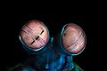
The eyes of mantis shrimps (here Odontodactylus scyllarus) are considered the best in the whole animal kingdom.
-
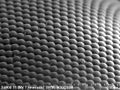
An image of a house fly compound eye surface by using scanning electron microscope
-
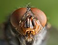
Arthropods such as this bluebottle fly have compound eyes
-
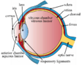
The structures of the eye labelled


See also
 In Spanish: Ojo para niños
