
Image: Anatomy of the cat (1991) (18195201631)

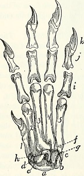
Description: Title: Anatomy of the cat Identifier: anatomyofcatrje00reig (find matches) Year: 1991 (1990s) Authors: Reighard, Jacob Ellsworth, 1861-1942; Jennings, H. S. (Herbert Spencer), 1868-1947 Subjects: Cats; Mammals Publisher: (Austin, TX) : BookLab, Inc. Contributing Library: American Museum of Natural History Library Digitizing Sponsor: Biodiversity Heritage Library View Book Page: Book Viewer About This Book: Catalog Entry View All Images: All Images From Book Click here to view book online to see this illustration in context in a browseable online version of this book. Text Appearing Before Image: 70 THE SKELETON OF THE CAT. ScapJioliinar Bone. Os scapJiobinaris (Fig. 51, a).—The scapholunar is a quadrangular bone with the ventroradial angle produced into a blunt process. Its proximal surface is smooth and articulates with the distal end of the radius. The distal end is marked by oblique ridges and articulates with the unci- form, OS magnum, trapezoid, and trapezium. The ulnar surface articulates with the cuneiform, and the dorsal surface of the ventroradial process with the radial sesamoid. Ciineiform Bone. (Os triqnetriim BNA) (Fig. 51, b).— The cuneiform bone has the form of a flattened pyramid. Its base articulates with the unciform, its proximoulnar surface with the pisiform except at its dorsal margin, where it articulates with the styloid process of the ulna. On its proximoradial sur- face is a smooth facet for articulation with the scapholunar. Pisiform Bone. Os pisiforme (Fig. 51, c).—The pisiform bone is about twice as long as broad, with enlarged ends. Its dorsal end articu- lates with the cuneiform, and on its proximal surface, separated from the above by a smooth ridge, is a smooth facet for articulation with the styloid process of the ulna. Unciform Bone. (Os hamatnin BNA) (Fig. 51, g).—The unciform is a wedge-shaped bone with the apex CARPUS, AND PuALANGEs, of the wcdgc dlrcctcd proximad, and Dorsal Surface. smooth for articulation with the a, scapholunar bone ; l>, . r -^ 1 cuneiform; ^, pisiform;^, trape- scapholunar. By a part of its ulnar zium; e, trapezoid; /, os mag- gurface it articulates with the cunei- num; g, unciform; h, radial i , . i- i r -in sesamoid;/.proximalphalanges; form, and by its radial Surface Wltll tlie J, second phalanges; /', distal ^^ niat^num. Its distal end articulates phalanges; I, 2, 3, 4, 5. meta- . , ^ ^ ,, , cr.i ^ 1 carpals in order from the radial with the fourth and httll metacarpals. ^'^^' Os magnnni. (Os capitat7im BNA) (Fig. 51, f). The os magnum may be described as an Text Appearing After Image: Fig. 51. — Cari'US, Meta- Note About Images Please note that these images are extracted from scanned page images that may have been digitally enhanced for readability - coloration and appearance of these illustrations may not perfectly resemble the original work.
Title: Anatomy of the cat (1991) (18195201631)
Credit: https://www.flickr.com/photos/internetarchivebookimages/18195201631/ Source book page: https://archive.org/stream/anatomyofcatrje00reig/#page/n99/mode/1up
Author: Internet Archive Book Images
Permission: At the time of upload, the image license was automatically confirmed using the Flickr API. For more information see Flickr API detail.
Usage Terms: No known copyright restrictions
License: No restrictions
License Link: https://www.flickr.com/commons/usage/
Attribution Required?: No
Image usage
The following page links to this image:

_(18195201631).jpg)