
Image: Antibody Opsonization
Size of this PNG preview of this SVG file: 512 × 339 pixels. Other resolution: 320 × 212 pixels.


Original image (SVG file, nominally 512 × 339 pixels, file size: 396 KB)

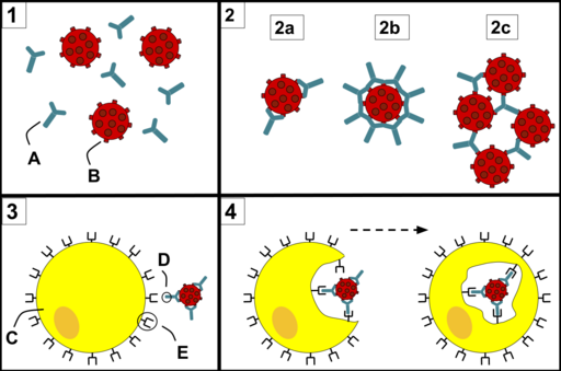
Description: 1) Antibodies (A) and pathogens (B) free roam in the blood. 2) The antibodies bind to pathogens, and can do so in different formations such as: opsonization (2a), neutralisation (2b), and agglutination (2c). 3) A phagocyte (C) approaches the pathogen, and the Fc region (D) of the antibody binds to one of the Fc receptors (E) of the phagocyte. 4) Phagocytosis occurs as the pathogen is ingested.
Author: Maher33
Usage Terms: Creative Commons Attribution-Share Alike 3.0
License: CC-BY-SA-3.0
License Link: http://creativecommons.org/licenses/by-sa/3.0/
Attribution Required?: Yes
Image usage
The following page links to this image:

All content from Kiddle encyclopedia articles (including the article images and facts) can be freely used under Attribution-ShareAlike license, unless stated otherwise.
