
Image: Bacteria-3D-Double-Helix

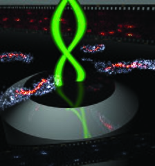
Description: This image shows 3D super-resolution imaging of Caulobacter crescentus bacteria cell surfaces (gray) and a labeled protein (CreS, orange-red) obtained using the double-helix single-molecule active control microscopy technique. For details, see Matthew D. Lew*, Steven F. Lee*, Jerod L. Ptacin, Marissa K. Lee, Robert J. Twieg, Lucy Shapiro, and W. E. Moerner (*equal contributions), “Three-dimensional super-resolution co-localization of intracellular protein superstructures and the cell surface in live Caulobacter crescentus,” Proc. Nat. Acad. Sci. (USA) 108, E1102-E1110 (2011) and 108, 18577-18578 (2011), DOI:10.1073/pnas.1114444108
Title: Bacteria-3D-Double-Helix
Credit: Own work
Author: WeoMoe
Usage Terms: Creative Commons Attribution-Share Alike 4.0
License: CC BY-SA 4.0
License Link: https://creativecommons.org/licenses/by-sa/4.0
Attribution Required?: Yes
Image usage
The following page links to this image:
