
Image: Birefringence microscopy of pseudogout, annotated

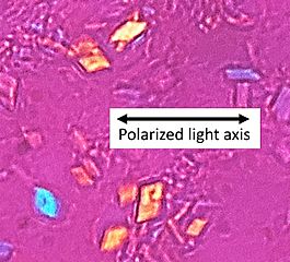
Description: Microscopy with polarized light of tissue by a metatarsal joint, showing crystals whereof some (one annotated) have rhomboid shape and weak positive nirefringence, consistent with calcium pyrophosphate dihydrate crystal deposition disease (pseudogout).
Title: Birefringence microscopy of pseudogout, annotated
Credit: Own work
Author: Mikael Häggström, M.D. - Author info - Reusing images - Conflicts of interest: None Mikael Häggström Consent note: Consent from the patient or patient's relatives is regarded as redundant, because of absence of identifiable features (List of HIPAA identifiers) in the media and case information (See also HIPAA case reports guidance).
Usage Terms: Creative Commons Zero, Public Domain Dedication
License: CC0
License Link: http://creativecommons.org/publicdomain/zero/1.0/deed.en
Attribution Required?: No
Image usage
The following page links to this image:
