
Image: CT scan showing liver and kidney
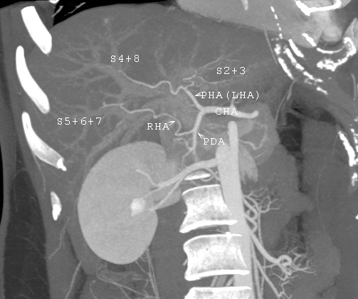
Description: A case referred for living donor evaluation. The scan is performed by a multi-detector row computed tomography. The post-processing is done by the interpreting radiologist, Dr. I-Chen Tsai. The picture shows an unusual variation of hepatic artery. The left hepatic artery supplies not only left lobe but also segment 8. The anatomy makes right lobe donation impossible. Even used as left lobe or lateral segment donation, it would be very technically challenging in anastomosing the small arteries.
Author: Dr. I-Chen Tsai
Usage Terms: Creative Commons Attribution-Share Alike 3.0
License: CC-BY-SA-3.0
License Link: http://creativecommons.org/licenses/by-sa/3.0/
Attribution Required?: Yes
Image usage
The following image is a duplicate of this image (more details):
The following page links to this image:
