
Image: Candidatus Prometheoarchaeum syntrophicum SEM Cryo
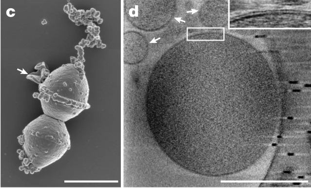
Description: MK-D1 cells dividing under SEM (c). Cryo-electron tomography image of a single cell (d). White arrows indicate large membrane vesicles. Scale bar = 1 μm (c) and 500 μm (d)
Title: Candidatus Prometheoarchaeum syntrophicum SEM Cryo
Credit: Isolation of an archaeon at the prokaryote–eukaryote interface. Nature 577, 519–525 (2020). https://doi.org/10.1038/s41586-019-1916-6
Author: Hiroyuki Imachi, Masaru K. Nobu,Nozomi Nakahara,Yuki Morono, Miyuki Ogawara, Yoshihiro Takaki, Yoshinori Takano, Katsuyuki Uematsu, Tetsuro Ikuta, Motoo Ito, Yohei Matsui, Masayuki Miyazaki, Kazuyoshi Murata, Yumi Saito, Sanae Sakai, Chihong Song, Eiji Tasumi, Yuko Yamanaka, Takashi Yamaguchi, Yoichi Kamagata, Hideyuki Tamaki, and Ken Takai
Usage Terms: Creative Commons Attribution-Share Alike 4.0
License: CC BY-SA 4.0
License Link: https://creativecommons.org/licenses/by-sa/4.0
Attribution Required?: Yes
Image usage
The following page links to this image:
