
Image: Elements of the comparative anatomy of vertebrates (1886) (21057027940)
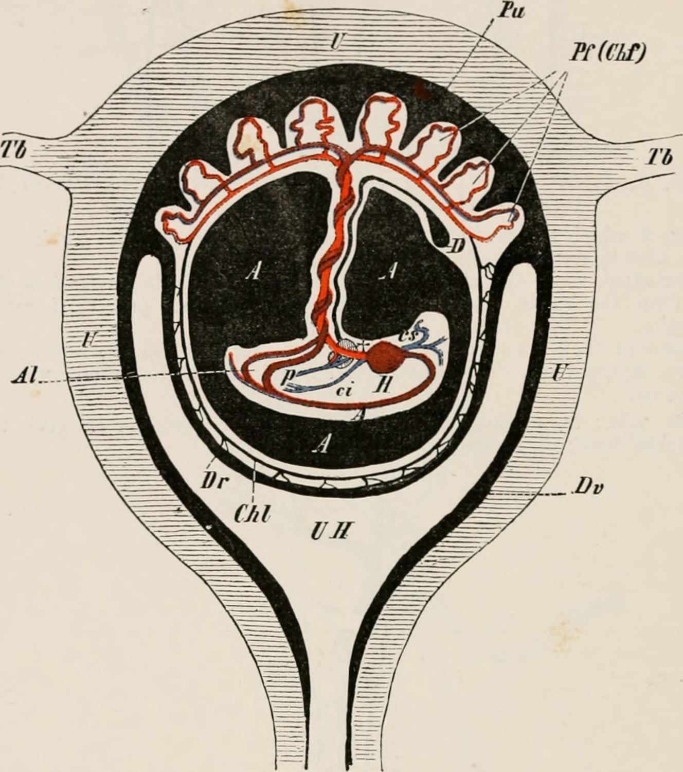
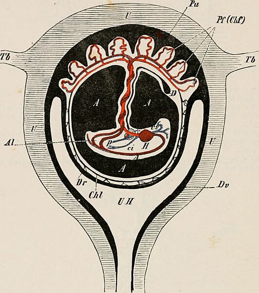
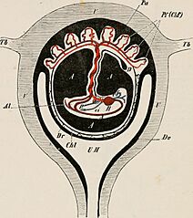
Description: Title: Elements of the comparative anatomy of vertebrates Identifier: elementsofcompar00wied (find matches) Year: 1886 (1880s) Authors: Wiedersheim, Robert, 1848-1923; Parker, W. N. (William Newton), d. 1923 Subjects: Anatomy, Comparative; Vertebrates -- Anatomy Publisher: London, New York, Macmillan Contributing Library: MBLWHOI Library Digitizing Sponsor: MBLWHOI Library View Book Page: Book Viewer About This Book: Catalog Entry View All Images: All Images From Book Click here to view book online to see this illustration in context in a browseable online version of this book. Text Appearing Before Image: 276 COMPARATIVE ANATOMY. definitive urinary bladder and urethra. Indications of the point of exit of the allantois and vitcllo-intestinal duct (umbilical cord) from the body-cavity can be seen in the adult at the navel, or umbilicus, Avhich represents the last point at which the body-walls become united. PC(CLf) Text Appearing After Image: FIG. 220.—DIAGRAMMATIC SECTION THROUGH THE HUMAN GUAVID UTERUS. U, uterus ; T/>, Tb, Fallopian tubes ; UIT, uterine cavity ; Dv, cleeidua vera, which at Pu passes into the uterine portion of the placenta ; 7V, decidna rellcxa ; Pf, f<etal portion of the placenta (chorion frondosuni, Clif); Chi, chorion Iteve ; A, .1, the cavity of the anniion filled with lluid : in the interior of the amnion is seen the embryo suspended by the twisted umbilical cord ; //, heart ; A, ami a ; cs, precaval ; <•!, postcaval; p, portal vein ; At, allantoic (umbilical) artcrio : f, the liver, perforated by the umbilical vein ; D, the remains of the yolk-sac (umbilical vesicle). The branchial vessels never become functional as such, in any period of development either in Mammalia or Suuropsida, but those which persist give rise to important vascular trunks of the neck, head (carotids), upper extremity (subclavian), and lungs (pulmonary artery), and also to the roots of the aorta, one or both of which m;iy remain (comp. Fig. 221, A to D). Note About Images Please note that these images are extracted from scanned page images that may have been digitally enhanced for readability - coloration and appearance of these illustrations may not perfectly resemble the original work.
Title: Elements of the comparative anatomy of vertebrates (1886) (21057027940)
Credit: https://www.flickr.com/photos/internetarchivebookimages/21057027940/ Source book page: https://archive.org/stream/elementsofcompar00wied/#page/n307/mode/1up
Author: Internet Archive Book Images
Permission: At the time of upload, the image license was automatically confirmed using the Flickr API. For more information see Flickr API detail.
Usage Terms: No known copyright restrictions
License: No restrictions
License Link: https://www.flickr.com/commons/usage/
Attribution Required?: No
Image usage
There are no pages that link to this image.

_(21057027940).jpg)