
Image: Enterobacteria phage T2 transmission electron micrograph

Size of this preview: 600 × 600 pixels. Other resolutions: 240 × 240 pixels | 2,021 × 2,021 pixels.
Original image (2,021 × 2,021 pixels, file size: 1.1 MB, MIME type: image/jpeg)
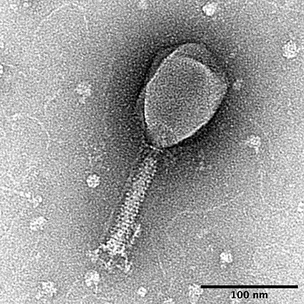
Description: T2 phage was prepared from Escherichia coli lysate by PEG precipitation then negatively stained with uranyl formate. Micrograph was taken with an FEI Tecnai transmission electron microscope operating at 120 keV. Cellular debris, nucleic acid, and possibly dissociated tail fibres are visible around the phage. Scale bar, 100 nm.
Title: Enterobacteria phage T2 transmission electron micrograph
Credit: Own work
Author: SnaxMikn
Usage Terms: Creative Commons Attribution-Share Alike 4.0
License: CC BY-SA 4.0
License Link: https://creativecommons.org/licenses/by-sa/4.0
Attribution Required?: Yes
Image usage
The following page links to this image:

All content from Kiddle encyclopedia articles (including the article images and facts) can be freely used under Attribution-ShareAlike license, unless stated otherwise.