
Image: Powell2004Fig1A
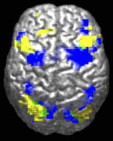
Description: fMRI-derived image of difference between brains of autistic and control groups. Legend reads "Activation during visuomotor coordination: Autism Group [yellow], Control Group [Blue], Overlap (both groups) [green]". for Laurent Mottron*, "Even researchers who study autism can display a negative bias against people with the condition. For instance, researchers performing functional magnetic resonance imaging (fMRI) scans systematically report changes in the activation of some brain regions as deficits in the autistic group — rather than evidence simply of their alternative, yet sometimes successful, brain organization". * Laurent Mottron, Changing perceptions: The power of autism ; Nature 479, 33–35 (03 November 2011) ; doi:10.1038/479033a En ligne : 2011-11-02
Title: Powell2004Fig1A
Credit: Figure 1A of: Powell K (2004). "Opening a window to the autistic brain". PLoS Biol 2 (8): E267. DOI:10.1371/journal.pbio.0020267. PMID 15314667. PMC: 509312.
Author: Ralph-Axel Müller
Permission: CCAL 2.5[1]
Usage Terms: Creative Commons Attribution 2.5
License: CC BY 2.5
License Link: http://creativecommons.org/licenses/by/2.5
Attribution Required?: Yes
Image usage
There are no pages that link to this image.
