
Image: Rheumatoid arthritis ultrasound MRI MCP joint ar1904-2

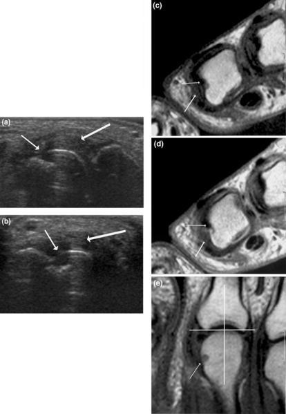
Description: Signs of destruction and inflammation on ultrasonography and MRI in second metacarpophalangeal joint: established RA. Thin arrows indicate an erosive change; thick arrows indicate synovitis. Ultrasonography in the (a) longitudinal and (b) the transverse planes shows both signs of destruction (grade 2) and inflammation (grade 3). Axial T1-weighted magnetic resonance images were obtained (c) before and (d) after contrast administration (grade 3 synovitis). Additionally, a coronal T1-weighted magnetic resonance image (e) before contrast administration visualizes the same bone erosion as shown in panels c and d. The coronal magnetic resonance image of the second metacarpophalangeal joint (panel e) is additionally covered by a grid illustrating division of the assessed joints into quadrants: proximal radial, proximal ulnar, distal radial and distal ulnar. MRI, magnetic resonance imaging; RA, rheumatoid arthritis.
Title: Rheumatoid arthritis ultrasound MRI MCP joint ar1904-2
Credit: Ultrasonography of the metacarpophalangeal and proximal interphalangeal joints in rheumatoid arthritis: a comparison with magnetic resonance imaging, conventional radiography and clinical examination. Arthritis Research & Therapy 2006, 8:R52. doi:10.1186/ar1904
Author: Marcin Szkudlarek, Mette Klarlund, Eva Narvestad, Michel Court-Payen, Charlotte Strandberg, Karl E Jensen, Henrik S Thomsen and Mikkel Østergaard.
Usage Terms: Creative Commons Attribution 2.0
License: CC BY 2.0
License Link: http://creativecommons.org/licenses/by/2.0
Attribution Required?: Yes
Image usage
There are no pages that link to this image.
