
Image: Richtersius coronifer in active and tun states

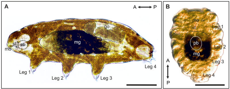
Description: Light microscopy of A. active, hydrated animal (lateral view) and B. a tun (ventral view) showing the rearrangement of major anatomical structures during tun formation. Note the compact body shape of the tun. Dashed circles indicate areas of the midgut (mg), gonads (go) and pharyngeal bulb (pb), respectively. The degree of longitudinal contraction is ultimately limited by the length of the rigid stylets (st). The pharyngeal bulb is for the most part repositioned in the dorsomedian plane. A↔P, anterior-posterior axis; br, brain; gI-gIV, ventral ganglia; mo, mouth. Scale bars = 100 μm. doi:10.1371/journal.pone.0085091.g001
Author: Halberg KA, Jørgensen A, Møbjerg N
Usage Terms: Creative Commons Attribution-Share Alike 3.0
License: CC-BY-SA-3.0
License Link: http://creativecommons.org/licenses/by-sa/3.0/
Attribution Required?: Yes
Image usage
The following page links to this image:
