
Image: Synapsin and CamK2 positive neurons

Size of this preview: 741 × 600 pixels. Other resolutions: 297 × 240 pixels | 1,180 × 955 pixels.
Original image (1,180 × 955 pixels, file size: 1.82 MB, MIME type: image/png)
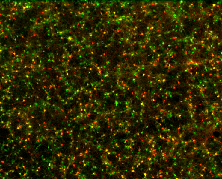
Description: Fluorescence microscopy picture of cortical neurons in a primary culture from E18 rat, grown in supplemented Neurobasal Medium. On DIV 2, the cells were transduced with two adeno-associated viruses [AAV9.hSyn.TurboRFP and AAV9.CamKII0.4.eGFP] that express red and green fluorescent proteins under the controls of the Synapsind and the CamKII promoters. Picture is taken DIV 7.
Title: Synapsin and CamK2 positive neurons
Credit: Own work
Author: ManuelSchottdorf
Usage Terms: Creative Commons Attribution-Share Alike 4.0
License: CC BY-SA 4.0
License Link: https://creativecommons.org/licenses/by-sa/4.0
Attribution Required?: Yes
Image usage
The following page links to this image:

All content from Kiddle encyclopedia articles (including the article images and facts) can be freely used under Attribution-ShareAlike license, unless stated otherwise.