
Image: Typhloesus interpretation 2022
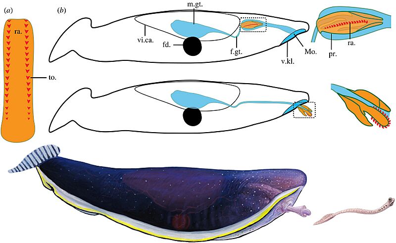
Description: Figure 2. Typhloesus wellsi: anatomical schematic diagrams and artistic reconstruction. (a) Interpretative reconstruction of the radula fully outstretched as seen from above, anterior to the top, showing two main rows of lateral teeth (red triangles) decreasing in size towards the rear; (b) interpretative sagittal sections of the body showing the gut system (blue) with a blind gut and the proboscis with the radula complex (orange) in a fully inverted (top) and everted (bottom) position. Framed areas, close-ups of anterior region of the proboscis; (c) artistic representation of Typhloesus wellsi in the process of catching its conodont prey using its everted proboscis and radula. Drawing by Joschua Knüppe © Royal Ontario Museum. fd., ferrodiscus; m.gt., midgut; mo., mouth; ra., radula; pr., proboscis; vi.ca., visceral capsule.
Author: Simon Conway Morris and Jean-Bernard Caron
Usage Terms: Creative Commons Attribution-Share Alike 3.0
License: CC-BY-SA-3.0
License Link: http://creativecommons.org/licenses/by-sa/3.0/
Attribution Required?: Yes
Image usage
The following page links to this image:
