Radiography facts for kids

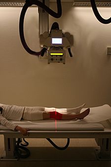
Radiography of the knee in a modern X-ray machine.
|
Radiography is a special way to look inside the human body or other objects without cutting them open. It uses X-rays, which are a type of energy similar to light, but you can't see them. X-rays can pass through many things that light cannot.
To create an image, an X-ray machine sends a beam of X-rays towards the object, like a part of your body. Different parts of the body, like bones or muscles, absorb different amounts of these X-rays. For example, bones are very dense, so they absorb a lot of X-rays. Soft tissues, like muscles, absorb less.
The X-rays that pass through the object are then caught by a special detector. This detector can be like a photographic film or a digital sensor. The detector then creates a flat, 2D picture that shows the inside of the object. This picture helps doctors see things like broken bones or other problems.
How X-rays Help Us See Inside
X-rays are a powerful tool for doctors. They help them understand what is happening inside your body. For example, if you break an arm, an X-ray can clearly show the broken bone. This helps the doctor know how to fix it.
X-rays are also used to check for other health issues. They can help find problems in your lungs, teeth, or other areas. It's a quick and painless way to get important information.
Advanced Imaging: CT Scans
Sometimes, doctors need an even more detailed look inside the body. This is where a more advanced technique called tomography comes in. A common type of tomography is called a CT scan, which stands for Computer Tomography.
Instead of just one flat picture, a CT scan takes many X-ray pictures from different angles around the body. Think of it like taking many "slices" of the body. A powerful computer then puts all these slices together. This creates a detailed 3D picture of the inside. This 3D view helps doctors see organs, bones, and tissues much more clearly. It can show problems that might be hidden in a regular 2D X-ray.
Radiography, including X-rays and CT scans, is used in many ways. It helps doctors diagnose and treat illnesses. It is also used in industries to check materials or products for hidden flaws.
Images for kids
-
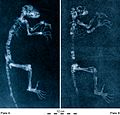
Radiography can also be used in paleontology, like for these X-rays of the Darwinius fossil Ida.
-
A plain X-ray of the elbow
-
1897 sciagraph (X-ray photograph) of Pelophylax lessonae (then Rana Esculenta), from James Green & James H. Gardiner's "Sciagraphs of British Batrachians and Reptiles"
See also
 In Spanish: Radiografía para niños


