History of computed tomography facts for kids
A CT scan (which stands for Computed Tomography scan) is a special type of X-ray. It creates detailed pictures of the inside of your body. Think of it like taking many X-ray pictures from different angles. Then, a computer puts them together to make a 3D image. This helps doctors see bones, organs, and soft tissues clearly.
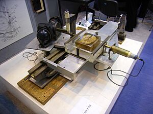
The idea for CT scans started a long time ago. In the early 1900s, an Italian doctor named Alessandro Vallebona came up with "stratigrafia." This was a way to see a single "slice" of the body using X-ray film. Later, in the 1930s, Dr. Bernard George Ziedses des Plantes made this method more practical.
In 1963, an American doctor named William H. Oldendorf got a patent for a device that could look inside objects using X-rays. But the first time a CT scan was used on a patient was in 1971. This happened with a machine invented by Sir Godfrey Hounsfield.
Contents
How CT Scans Work (The Math Behind It)
The basic idea for CT scans comes from math. Back in 1917, a mathematician named Johann Radon showed that you could rebuild a picture from many different "projections" or views. Imagine looking at an object from every side. Radon's math showed how to put all those views together to create the full object.
Later, in 1937, Stefan Kaczmarz developed a way to solve many math problems at once. This method, along with work by Allan McLeod Cormack, helped create the "algebraic reconstruction technique." This is the computer method Godfrey Hounsfield used to build the first CT scanner. It helped turn the X-ray data into clear images.
In 1959, Dr. William Oldendorf from UCLA had an idea. He thought about scanning a head with X-rays. He wanted to rebuild the image to see the different parts inside. In 1961, he built a simple machine. It had an X-ray source and a detector that spun around an object. This machine could take a picture of a nail even if other nails were blocking it from a single angle. His work helped lay the groundwork for modern CT.
Early CT Scanners
CT technology has come a long way. Early machines were slow and produced less clear images. Today's scanners are much faster and show amazing detail. This helps doctors diagnose problems and perform medical procedures more accurately.
The EMI Scanner
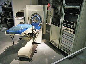
The first CT scanner that was actually used in hospitals was invented by Sir Godfrey Hounsfield. He worked at EMI Central Research Laboratories in the United Kingdom. Hounsfield came up with his idea in 1967.
The very first EMI-Scanner was put in a hospital in Wimbledon, England. On October 1, 1971, the first patient had a brain scan using this machine. The invention was announced to the public in 1972.
The first machine in 1971 was quite slow. It took over 5 minutes to scan one "slice" of the brain. Then, it took another 2.5 hours for a large computer to process the images! This scanner used a thin X-ray beam and a single detector.
The first production model of the EMI-Scanner was only for brain scans. It took about 4 minutes to get the data for two slices. The computer then took about 7 minutes to create each picture. Patients had to put their head in a special water-filled tank. This tank helped the X-rays pass through the skull better. The images were not very clear, made up of only 80x80 tiny squares (pixels).
In the U.S., the first EMI scanner was installed at the Mayo Clinic. Allan McLeod Cormack also worked on similar ideas independently. Both Hounsfield and Cormack won the Nobel Prize in Medicine in 1979 for their important work on CT.
The ACTA Scanner
The first CT system that could scan any part of the human body was called the ACTA scanner. ACTA stands for Automatic Computerized Transverse Axial. Dr. Robert S. Ledley designed this machine at Georgetown University.
This new machine was much faster than the EMI-Scanner. It had 30 detectors and could finish a scan in just nine quick movements. The ACTA scanner used a DEC PDP11/34 computer to control its parts and process the images.
A company called Pfizer bought the rights to make the ACTA scanner. They released their own version called the "200FS." This machine made much clearer images, with 256x256 pixels. It took about 20 seconds to get one image slice. This made it possible to scan the whole body, though patients still had to hold their breath during the scan. The EMI scanner couldn't do body scans because it was too slow.
The ACTA and 200FS machines required a lot of work from the operators. They would scan many slices and then process the images. These images were printed onto films. The raw data was saved on magnetic tapes because the computer didn't have enough storage for everything.
The DEC PDP11/34 computer was very important for these scanners. It controlled the scanning process and turned the raw data into final images. This computer worked with only 64 KB of memory and a small 5 MB hard disk.
Portable Scanners
Today, there are even portable CT scanners. These can be brought right to a patient's bedside. This means patients don't have to be moved to a special room for a scan. Some portable scanners are mainly used for head scans because of their size. They don't replace the big CT machines in hospitals, but they are very helpful in certain situations.
In 2008, Siemens introduced a new scanner that could take an image in less than 1 second. This speed is amazing! It's fast enough to get clear pictures of beating hearts and coronary arteries (the blood vessels around the heart).
Modern CT machines can also use a "spiral technique." The machine spins continuously while the patient moves through it. This quickly covers all the areas doctors want to see. This method also helps reduce the amount of X-ray exposure for the patient.
Photon-Counting Scanners
In 2021, a new type of scanner called a photon-counting scanner was approved. This scanner counts individual X-ray particles (photons) that pass through the patient. It can also tell the difference between their energy levels. This gives doctors even more detailed images. Plus, it uses less X-rays, which is safer for patients.
Older Techniques Replaced by CT
Before CT scans, doctors used other methods to see inside the body. CT scans replaced many of these older, less effective ways.
Focal Plane Tomography
Before CT, doctors used something called focal plane tomography. This method also tried to show a single "slice" of the body on an X-ray film. Alessandro Vallebona, the Italian radiologist, proposed this method early on.
The idea was to move the X-ray tube and the film at the same time but in opposite directions. This made the image of the "focal plane" (the slice you wanted to see) appear sharper. Images from other parts of the body would blur out. However, this blurring was only partly effective.
Over time, these mechanical methods got better. They could make sharper images and let doctors choose the thickness of the slice. More complex machines could move in different directions to blur out unwanted parts more effectively. But even with these improvements, focal plane tomography was not good at showing soft tissues.
With the rise of computers in the 1960s, scientists started working on computer-based ways to create tomographic images. This led to the development of the CT scan, which was much better at seeing soft tissues and provided clearer, more detailed images.
 | Tommie Smith |
 | Simone Manuel |
 | Shani Davis |
 | Simone Biles |
 | Alice Coachman |


