
Image: Chlamydomonas TEM 07

Size of this preview: 751 × 600 pixels. Other resolutions: 301 × 240 pixels | 1,800 × 1,438 pixels.
Original image (1,800 × 1,438 pixels, file size: 878 KB, MIME type: image/jpeg)
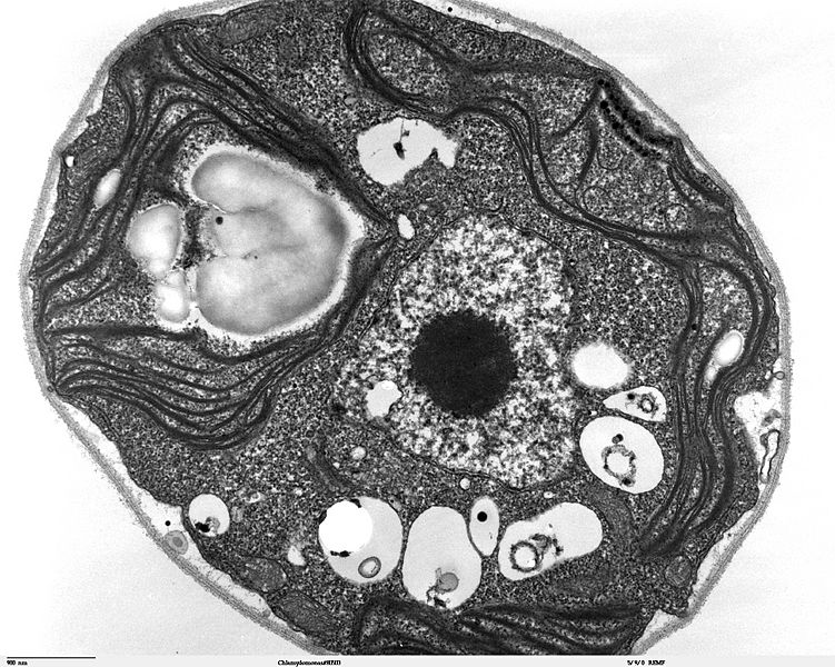
Description: Transmission electron microscope image, showing an example of green algae (Chlorophyta). Chlamydomanas reinhardtii is a unicellular flagellate used as a model system in molecular genetics work and flagellar motility studies. This image of a thin section through a whole Chlamydomonas, shows the nucleus, chloroplast, starch grains, vacuoles, mitochondria, eye spot, and the cell wall.
Title: Chlamydomonas TEM 07
Credit: Source and public domain notice at http://remf.dartmouth.edu/imagesindex.html
Author: Dartmouth Electron Microscope Facility, Dartmouth College
Permission: Released into the public domain
Usage Terms: Public domain
License: Public domain
Attribution Required?: No
Image usage
The following page links to this image:

All content from Kiddle encyclopedia articles (including the article images and facts) can be freely used under Attribution-ShareAlike license, unless stated otherwise.