
Histology facts for kids
Histology is the study of tiny parts of living things. It looks at cells and tissues from plants and animals using powerful microscopes. Think of it as looking at the building blocks of life up close!
Histology is a big part of biology and medicine. It helps scientists and doctors understand how bodies work and what happens when they get sick. To study these tiny parts, tissues need to be specially prepared.
Contents
How Histology Works
Studying tissues under a microscope takes several steps. These steps make sure the tissues are preserved and easy to see. Here's a simple look at the process:
Keeping Tissues Safe
The first step is called fixing. This means using special chemicals to stop tissues from decaying. It's like freezing them in time! This helps keep the cells and their tiny parts, like the nucleus or mitochondria, looking just as they were. A common chemical used for this is called formalin.
Making a Tissue Block
After fixing, the tissue is put into a block of paraffin wax. This step is called embedding. The wax holds the tissue firmly in place. It also protects the tissue so it can be cut very thinly without falling apart.
Cutting Thin Slices
Next, the wax block with the tissue inside is cut into super-thin slices. This is done using a special machine called a microtome, which is like a very precise slicer. Each slice is thinner than a human hair! These tiny slices are then carefully placed onto a glass microscope slide.
Adding Color to Tissues
The slices are almost invisible on the slide. So, the next step is staining. This means adding special dyes to the tissue. These dyes make different parts of the cells and tissues stand out with different colors. This makes them much easier to see and study under a microscope. There are many types of stains, each with its own use.
Common Stains
One of the most popular stains is called Haematoxylin and eosin (often shortened to H&E).
- Haematoxylin makes the nuclei (the control center of the cell) look blue or purple.
- Eosin makes the cytoplasm (the jelly-like substance around the nucleus) look pink or red.
This combination helps scientists see the basic structure of cells and tissues clearly.
Another important stain uses silver nitrate. A scientist named Camillo Golgi first used a silver stain to see nerve cells in amazing detail. Later, Santiago Ramón y Cajal used this idea to map out the complex structure of the brain.
Modern Ways to Study Tissues
Today, scientists use more than just light microscopes. They also use very powerful electron microscopes.
- Electron microscopes can see things that are too small for light microscopes, like tiny viruses. They have their own special ways of preparing tissues.
Scientists also use advanced staining methods.
- Some methods use immunology, which means they use special antibodies that stick to specific molecules in the tissue.
- Others use radioactive labels or fluorescent stains. These stains glow brightly, even if only a tiny part of a cell is stained. This technique is called immunofluorescence. These modern tools help scientists find and study very specific parts of cells and tissues.
Images for kids
-
A stained tissue sample on a microscope slide ready for viewing.
-
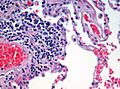
A close-up view of human lung tissue, stained with H&E, as seen under a microscope.
-
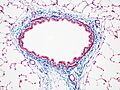
A rat airway stained with Masson's trichrome. Connective tissue is blue, nuclei are dark red/purple, and cytoplasm is red/pink.
-
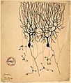
A drawing of Purkinje cells from a pigeon's brain by Santiago Ramón y Cajal (1899).
-
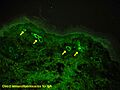
A view of human skin showing IgA deposits (yellow arrows) using a special fluorescent stain.
-
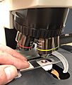
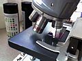
A tissue sample being placed on the stage of a light microscope.
-
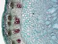
A cross-section of a plant stem (garlic mustard) under a microscope.
-
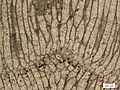
A thin slice of a fossilized invertebrate called a bryozoan.
-
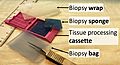
Items used to collect and send tissue samples for study.
-
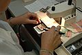
A tissue sample being embedded in warm paraffin wax.
-
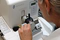
A machine called a microtome cutting very thin slices from a tissue block.
-
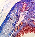
Masson's trichrome stain on a rat's trachea.
-
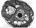
Green algae seen through a powerful Transmission electron microscope.
-
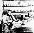
Santiago Ramón y Cajal working in his laboratory.


.jpg)


See also
 In Spanish: Histología para niños
