
Image: Journal.pone.0057573.g005 cropped
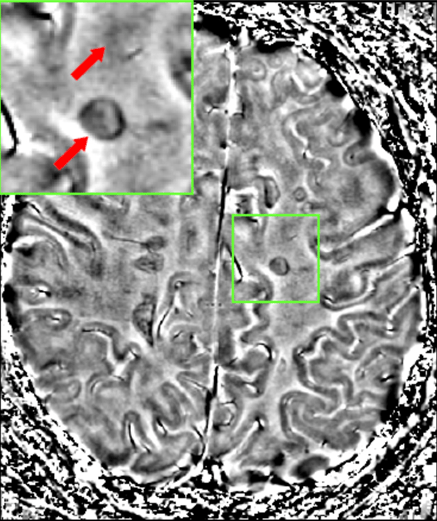
Description: Description in article for the original image: Figure 5. FLAIR (3T) and GRE phase (7T) images of a patient with active relapsing-remitting MS. FLAIR images show numerous white matter MS lesions of which 2 are magnified (inset, red arrows). Phase imaging at 7T phase/GRE reveals a hypointense ring corresponding with one lesion on FLAIR. The other lesion is not visible on 7T GRE (inset, arrows). After cropping, only the GRE phase brain scan (right of the original image) was left. Other info copied from article by uploader of file: Gradient-echo (GRE) phase imaging at ultra-highfield MRI is highly sensitive for iron. A number of GRE studies with MS patients and autoptic MS tissue, including our own have demonstrated that iron accumulates in white matter and cortical lesions.
Title: Journal.pone.0057573.g005 cropped
Credit: This file was derived from Journal.pone.0057573.g005.png:
Author: Journal.pone.0057573.g005.png: Veela Mehta, Wei Pei, Grant Yang, Suyang Li, Eashwar Swamy, Aaron Boster, Petra Schmalbrock, David Pitt derivative work: Garrondo
Usage Terms: Creative Commons Attribution 2.5
License: CC BY 2.5
License Link: http://creativecommons.org/licenses/by/2.5
Attribution Required?: Yes
Image usage
The following page links to this image:
