
Image: Liver reticulin
Size of this preview: 800 × 533 pixels. Other resolutions: 320 × 213 pixels | 4,272 × 2,848 pixels.
Original image (4,272 × 2,848 pixels, file size: 5.9 MB, MIME type: image/jpeg)
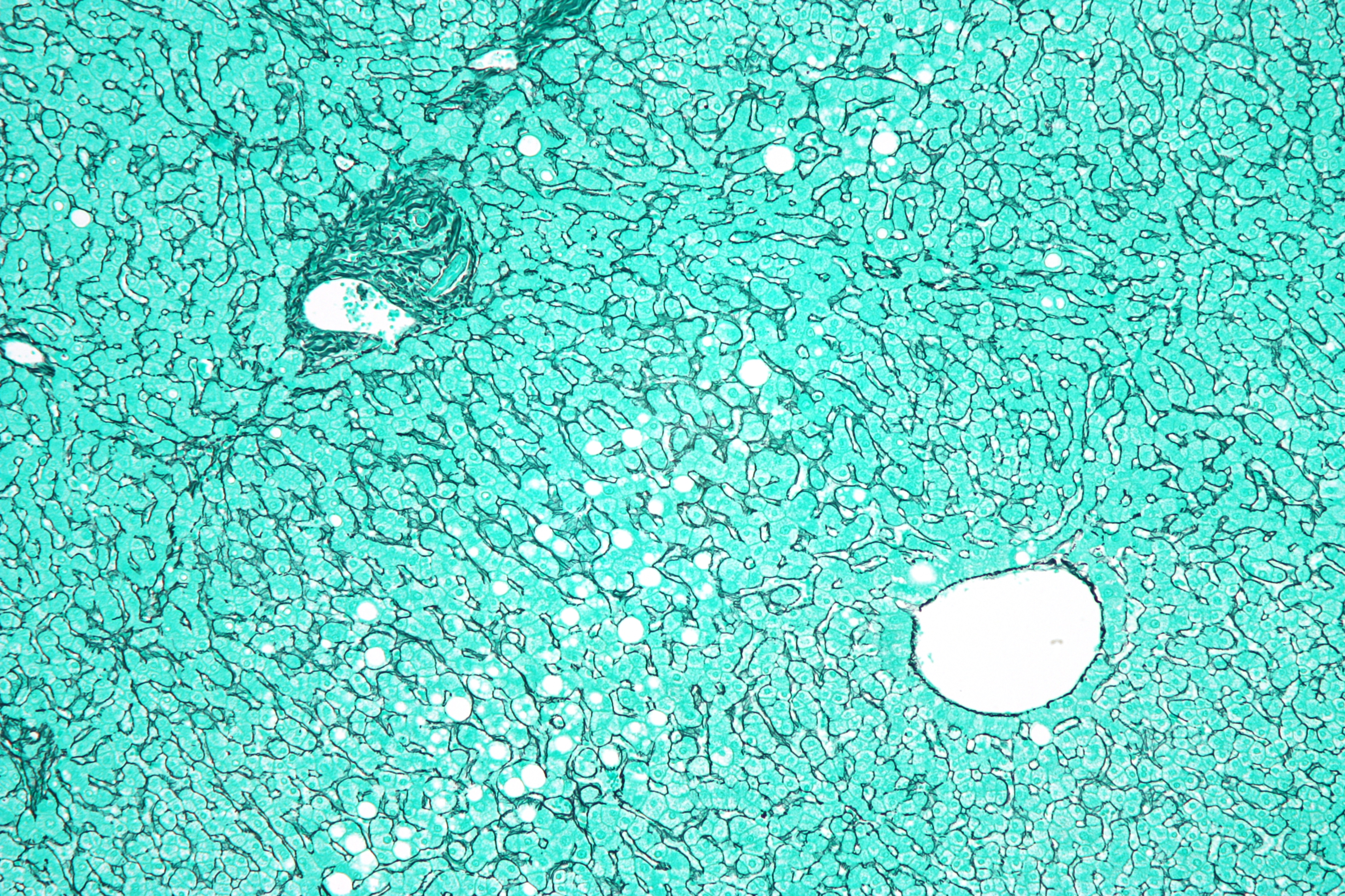
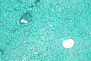
Description: Micrograph of the liver showing a portal tract and a nearby hepatic venule. Reticulin stain. The normal "hepatocyte plate" -- the strands of hepatocytes that characterise the morphology of the liver and are separated by the liver sinusoids -- is one or two cells thick, as shown in the image. If the hepatocyte plate is three or more cells, it is diagnostic of hepatocellular carcinoma, the most common form of cancer that arises in the liver.
Author: Nephron
Usage Terms: Creative Commons Attribution-Share Alike 3.0
License: CC-BY-SA-3.0
License Link: http://creativecommons.org/licenses/by-sa/3.0/
Attribution Required?: Yes
Image usage
The following page links to this image:

All content from Kiddle encyclopedia articles (including the article images and facts) can be freely used under Attribution-ShareAlike license, unless stated otherwise.