
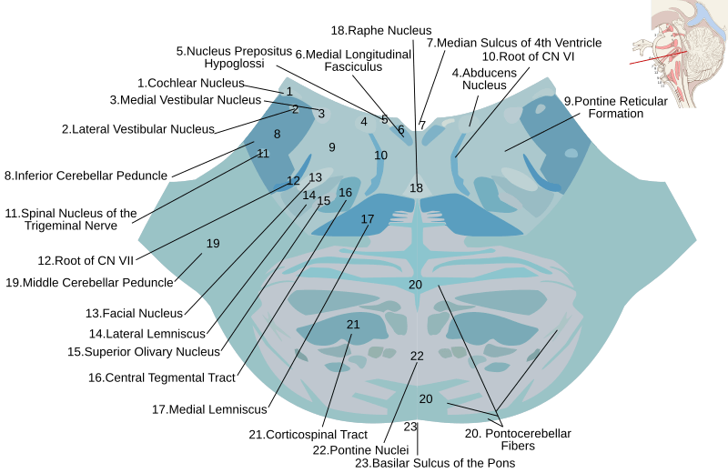
Image: Lower pons horizontal KB

Description: Diagram of a cross-section taken horizontally through the lower part of the pons of a human brainstem and stained with the Kluver-Barrera method. Due to the staining method, white matter (axons) appears blue and gray matter (cell bodies) appears light gray.
Notes:
Anterior is down, posterior is up. The spinal tract of the trigeminal nerve can sometimes be seen surrounding the spinal nucleus of the trigeminal nerve, but it was impossible to distingusih from the inferior cerebellar peduncle on the original slide. The spinal lemniscus (including the spinothalamic tract) and trapezoid body also could not be distinguished but probably lie anteromedially to the superior olivary nucleus, blending in with the central tegmental tract and medial lemniscus. The pontine reticular formation (#9) and pontine nuclei (#22) are large, diffuse structures.
Colors, shapes, and locations are based loosely on slide "05-BBS-09. Pons, Nu VII, Sup. Olive Kluver-Berrara" from the Bacus collection at Washington University in St. Louis School of Medicine, supplemented with information from Gado, Thomas A. Woolsey ; Joseph Hanaway ; Mokhtar H. (2003) The brain atlas a visual guide to the human central nervous system (2nd ed.), Hobok Wiley, pp. 146-147 ISBN: 0-471-43058-7. and several of the public domain images from Gray's anatomy on the Wikipedia Commons. Locater image illustrating point of section is File:Brain_stem_sagittal_section.svg by Patrick Lynch. Special thanks to Dr. Joel Price for his assistance in verifying structures.
Author: Marshall Strother User:mcstrother
Brain_stem_sagittal_section.svg: Patrick J. Lynch, medical illustrator
Usage Terms: Creative Commons Attribution-Share Alike 3.0
License: CC-BY-SA-3.0
License Link: http://creativecommons.org/licenses/by-sa/3.0/
Attribution Required?: Yes
Image usage
The following page links to this image:
