
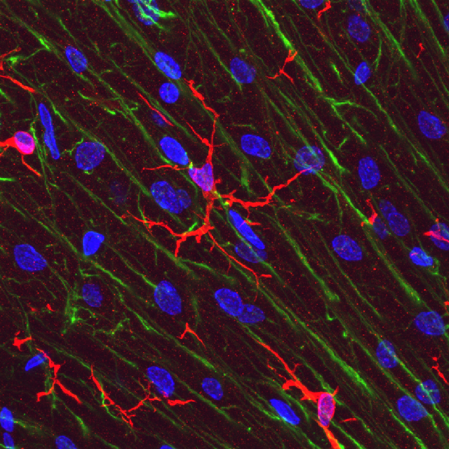
Image: Microglial cells (red) in rat cerebellar molecular layer
Size of this preview: 600 × 600 pixels. Other resolutions: 240 × 240 pixels | 919 × 919 pixels.
Original image (919 × 919 pixels, file size: 512 KB, MIME type: image/jpeg)
Description: Rat cerebellar molecular layer stained with IBA-1 antibody which reveals microglial cells (red). Bergmann glia are revealed in green with antibody to GFAP. Nuclear DNA is shown in blue with DAPI stain. Antibodies and image generated by EnCor Biotechnology Inc.
Title: Microglial cells (red) in rat cerebellar molecular layer
Credit: Z-stack confocal image made with 100X lens on Olympus FV1000 microscope.
Author: GerryShaw
Usage Terms: Creative Commons Attribution-Share Alike 4.0
License: CC BY-SA 4.0
License Link: https://creativecommons.org/licenses/by-sa/4.0
Attribution Required?: Yes
Image usage
The following page links to this image:

All content from Kiddle encyclopedia articles (including the article images and facts) can be freely used under Attribution-ShareAlike license, unless stated otherwise.
_in_rat_cerebellar_molecular_layer.jpg)