Microglia facts for kids
Quick facts for kids Microglia |
|
|---|---|
 |
|
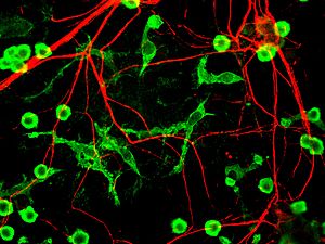
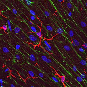
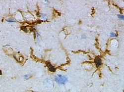
| Microglia in a resting state from a rat brain before injury. | |
 |
|
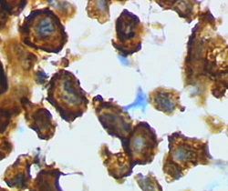
| Microglia/macrophage – an active form from a rat brain after injury. | |
| System | Central nervous system |
| Precursor | Primitive yolk-sac derived macrophage |
Microglia are special cells found all over your brain and spinal cord. They are a type of brain cell that makes up about 10-15% of all cells in your brain. Think of them as the brain's own tiny clean-up crew and security guards!
Microglia are the main part of your brain's immune system. They are always busy looking for problems. This includes finding unwanted clumps, damaged nerve cells, or even tiny germs that might get into your central nervous system (CNS). They are super sensitive to any small changes in the brain. This helps them react quickly to keep your brain healthy.
Your brain and spinal cord are protected by something called the blood–brain barrier. This barrier is made of special cells that stop most infections from getting into your delicate nervous system. But if germs do get in, microglia quickly jump into action. They work to reduce inflammation and destroy the germs before they can cause damage. Since there are not many antibodies from the rest of your body in the brain, microglia have to be able to recognize and fight off invaders all by themselves. They can even "eat" foreign bodies and show parts of them to other immune cells, like T-cells, to get more help.
Contents
How Microglia Change Their Shape
Microglial cells are very flexible. They can change their shape and how they act depending on what the brain needs. This ability to change helps them do many different jobs. It also means they can react very quickly to protect your brain without causing too much trouble to the immune system. Microglia change their form based on what they sense around them.
Resting Microglia: The Surveyors
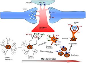
This type of microglia is often found throughout the brain and spinal cord when everything is healthy. They are called "resting" microglia, but they are actually very active! They have long, branching arms that are constantly moving. These arms survey the area around them. They are very sensitive to even tiny changes in the brain's condition.
Resting microglia do not "eat" other cells or release many immune molecules. Their job is to look for threats while keeping the brain balanced. They can quickly change into an active form if there is an injury or danger.
Active Microglia: The Responders
When microglia detect a problem, they become "reactive" or "activated." This term is better than "activated" because even "resting" microglia are active. A protein called Iba1 often shows up more when microglia are reactive, helping scientists see them.
Non-Eating Active Microglia
This is an early stage of microglia becoming fully active. They can be activated by things like chemicals that cause inflammation, damaged cells, or changes in potassium levels. When they activate, their arms get thicker and pull back. They also start to show special proteins and release molecules that cause more inflammation. These microglia might look "bushy" or like small blobs. They also multiply quickly to increase their numbers.
Eating Active Microglia
These are the most active form of microglia. They usually become large and blob-shaped. Besides causing inflammation and showing proteins to other immune cells, they can also "eat" foreign materials. These eating microglia travel to the injury site, swallow the bad stuff, and release chemicals to get more cells to help. They also work with other brain cells, like astrocytes, to fight off infection or inflammation. They try to do this with as little damage as possible to healthy brain cells.
Amoeboid Microglia: The Clean-up Crew
This blob-like shape allows microglia to move freely through brain tissue. This helps them act as "scavenger" cells. Amoeboid microglia can eat debris, but they do not play the same role in showing antigens or causing inflammation as the "active" microglia. These microglia are very common when the brain is developing. At this time, there is a lot of extra debris and dying cells that need to be removed. You can find many of them in areas like the "Fountains of Microglia" in the corpus callosum.
Gitter Cells: The Full Microglia
Gitter cells are what microglia become after they have eaten a lot of infectious material or cell debris. Once a microglia has eaten a certain amount, it cannot eat any more. This full cell is called a granular corpuscle because it looks "grainy." Scientists can look for gitter cells to see areas where the brain has healed after an infection.
Microglia Near Blood Vessels
Some microglia are named for where they are found:
- Perivascular microglia: These are found inside the walls of blood vessels. They do normal microglia jobs, but they are regularly replaced by new cells from the bone marrow. They also show special immune proteins no matter what is happening around them. These microglia are important for repairing damaged blood vessels.
- Juxtavascular microglia: These are found right next to the walls of blood vessels but not inside them. Like perivascular cells, they show special immune proteins even with low inflammation. But unlike perivascular cells, they are not replaced as often.
What Microglia Do
Microglial cells have many important jobs in the CNS. They help with the immune system and keep the brain balanced. Here are some of their main functions:
Cleaning Up the Brain
Each microglial cell constantly checks its own area. If it finds any foreign material, damaged cells, dying cells, or unwanted clumps, it will become active and "eat" the material. In this way, microglia act like "housekeepers," cleaning up random cell debris. When the brain is developing, microglia help remove dying nerve cells. They can also help trim connections between nerve cells.
Eating Bad Stuff (Phagocytosis)
The main job of microglia is to "eat" various materials. This includes cell debris, fats, and dying cells when the brain is healthy. When there's an infection, they eat invading viruses, bacteria, or other foreign materials. Once a microglia is "full," it stops eating and turns into a gitter cell.
Sending Signals
A big part of what microglia do is keep the brain balanced in healthy areas and cause inflammation in infected or damaged areas. They do this by sending out many different signals. These signals allow them to talk to other microglia, astrocytes, nerves, and other immune cells. For example, when activated, microglia can release more chemicals that activate other microglia, starting a chain reaction. Some signals cause nerve cells to die if they are infected, while others help recruit more immune cells to the site of injury.
Showing Antigens
Normally, microglia are not very good at showing "antigens" (parts of invaders) to other immune cells. But when they become active, they quickly get better at it. During inflammation, T-cells can cross the blood–brain barrier and connect directly with microglia to get these antigens. Once T-cells see the antigens, they go on to help fight the infection in many ways.
Releasing Harmful Chemicals
Microglia can also release chemicals that directly harm cells. They can produce hydrogen peroxide and nitric oxide, which can damage cells and lead to nerve cell death. They also release enzymes that break down proteins, causing direct cell damage. Some chemicals they release can even damage the protective coating around nerve fibers. These harmful chemicals are meant to destroy infected nerve cells, viruses, and bacteria. However, they can also cause damage to healthy brain cells. If inflammation lasts too long, it can lead to a lot of nerve damage as microglia try to destroy the infection.
Trimming Connections
Microglia can remove the branches from nerves near damaged tissue. This helps the brain regrow and rewire its connections. Microglia are also involved in trimming connections between nerve cells during brain development.
Helping Repair the Brain
After inflammation, microglia help the brain heal. They trim nerve branches, release chemicals that reduce inflammation, and help bring new nerve cells and astrocytes to the damaged area. They also form gitter cells. Without microglia, the brain would heal much slower. Recent studies show that microglia constantly check on nerve cells and can protect them, helping with repair after brain injury.
Why Microglia Are Important for Health
Microglia are the main immune cells of the central nervous system. They react to germs and injuries by changing their shape and moving to the problem area. There, they destroy germs and remove damaged cells. They also release chemicals that help guide the immune response. Microglia are also important for stopping inflammation once the danger is gone.
Scientists have studied how microglia can be harmful in brain diseases like Alzheimer's disease, Parkinson's disease, and Multiple sclerosis. They are also being studied in heart diseases, eye problems like glaucoma, and infections. There is growing evidence that problems with the immune system, including microglia, play a role in conditions like OCD and Tourette syndrome.
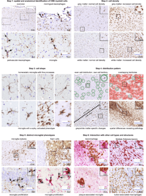
Because microglia react quickly to even small changes in the brain, they can be like sensors for brain problems. If there is a brain disease, the microglia will definitely change. So, studying microglia can be a helpful way to find and understand brain disorders. Scientists look at how many microglia there are, their shape, where they are found, and how they interact with other cells.
History of Discovering Microglia
Scientists first started seeing and describing different brain cells, including microglia, in the 1880s. Two scientists, Franz Nissl and William Ford Robertson, were among the first to describe microglial cells. Their staining methods showed that microglia were related to macrophages.
The first time someone noticed microglia becoming active and forming clusters was in 1897. Victor Babeş saw these cells while studying a case of rabies. He noticed them in many viral brain infections but did not know what they were.
A Spanish scientist named Santiago Ramón y Cajal talked about a "third element" (a cell type) in the brain, besides nerve cells and astrocytes. Then, around 1920, Pío del Río Hortega, one of Cajal's students, was the first to call these cells "microglia." He went on to describe how microglia react to brain injuries in 1927. In 1932, he noted the "fountains of microglia" in the corpus callosum and other areas. Rio Hortega is often called the "Father of Microglia" for all his research.
For many years, not much new was learned about microglia. Then, in 1988, Hickey and Kimura showed that microglia near blood vessels came from the bone marrow. They also found that these cells showed high levels of special proteins used for showing antigens. This confirmed what Pio Del Rio-Hortega had thought: that microglial cells worked like macrophages by "eating" things and showing antigens.
See also
 In Spanish: Microglía para niños
 | Leon Lynch |
 | Milton P. Webster |
 | Ferdinand Smith |


