
Bacteria facts for kids
Quick facts for kids Bacteria |
|
|---|---|
 |
|
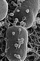
| Scanning electron micrograph of Escherichia coli rods | |
| Scientific classification |
|
| Domain: | Bacteria Woese, Kandler & Wheelis, 1990 |
Bacteria (say: bak-TEER-ee-uh) are super tiny living things. Most bacteria are so small you can only see them with a special microscope. They are some of the oldest and simplest living things on Earth.
Each bacterium is made of just one cell, making them a type of unicellular organism. They were among the very first forms of life to appear on our planet. There are probably more individual bacteria than any other type of living thing!
Most bacteria live in the ground or in water. But many also live inside or on the skin of other living things, including humans. Did you know there are about the same number of bacterial cells in your body as human cells?
Some bacteria can make us sick by causing diseases. But many others are helpful! For example, some help us digest food in our stomachs (gut flora). Others even help make yummy foods like cheese and yogurt in factories.
A German biologist named Ferdinand Cohn (1828–1898) was a founder of the study of bacteria, called bacteriology. He was the first to group bacteria based on how they looked.
Contents
What Do Bacteria Look Like?

Bacteria come in many different sizes and shapes. But they are usually at least ten times bigger than viruses. A typical bacterium is about 1 µm (one micrometer) across. This means a thousand bacteria lined up would only be one millimeter long! Scientists estimate there are about five nonillion (that's a 5 with 30 zeros!) bacteria on Earth.
Scientists often group bacteria by their shapes.
- Bacilli are shaped like rods.
- Cocci are shaped like tiny balls.
- Spirilla are spiral-shaped.
- Vibrio look like a comma or a boomerang.
Good Bacteria vs. Bad Bacteria
Some bacteria are helpful, but others can be harmful. The harmful ones are called pathogens.
How Harmful Bacteria Make Us Sick
Harmful bacteria can enter your body through the air, water, or food. Once inside, they can attach to or invade specific cells. This often happens in your breathing system, digestive system, or any open cut. There, they start to grow and spread. They use your body's food and nutrients to get energy and multiply. This process can make you feel sick.
For example, bacteria like Salmonella, Campylobacter, E. coli, and Listeria can cause food poisoning. This happens when these bacteria get into the food we eat, especially if it's old or hasn't been kept cold enough.
How to Stay Safe from Bad Bacteria

You can often avoid harmful bacteria by washing your hands regularly with soap and water. This washes away any germs you might have picked up from touching things.
If you scrape your knee, cleaning the wound with antiseptic helps stop bad bacteria from getting into your body.
Super Survivors: Extremophiles
Some bacteria are known as extremophiles. These are amazing bacteria that can live in very harsh conditions. For example, some can survive deep inside rocks, up to 580 meters below the sea floor! One researcher said, "You can find microbes everywhere — they're extremely adaptable to conditions, and survive wherever they are."
Amazing Facts About Bacteria

- There are four main types of germs: bacteria, viruses, fungi, and protozoa.
- Most of the bacteria in your body live in your gut. But they can be found almost anywhere on or inside you!
- Always flush the toilet with the lid down! If the lid is up, flushing can send a cloud of tiny bacteria into the air from the toilet bowl.
- Bacteria are all over household items like TV remote controls, phones, and cutting boards. A kitchen sponge can have up to forty-five billion microbes per square centimeter! It's a good idea to replace your kitchen sponges often.
- One reason we cook food is to heat it up and kill off any harmful bacteria.
- Some bacteria are important for the environment. They help break down dead plants and animals.
- Scientists have found a type of bacteria that can eat plastic! This bacteria eats polyurethane, which is hard to recycle and breaks down very slowly. This discovery could be a big step in reducing plastic waste on Earth.
Related Pages
Images for kids
-
Rod-shaped Bacillus subtilis
-
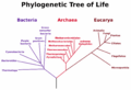
Phylogenetic tree of Bacteria, Archaea and Eucarya. The vertical line at bottom represents the last universal common ancestor.
-

An electron micrograph of Halothiobacillus neapolitanus cells with carboxysomes inside, with arrows highlighting visible carboxysomes. Scale bars indicate 100 nm.
-
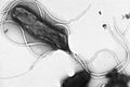
Helicobacter pylori electron micrograph, showing multiple flagella on the cell surface
-
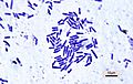
Bacillus anthracis (stained purple) growing in cerebrospinal fluid
-
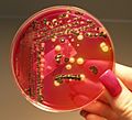
A culture of Salmonella
-
A colony of Escherichia coli
-
Helium ion microscopy image showing T4 phage infecting E. coli. Some of the attached phage have contracted tails indicating that they have injected their DNA into the host. The bacterial cells are ~ 0.5 µm wide.
-
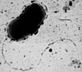
Transmission electron micrograph of Desulfovibrio vulgaris showing a single flagellum at one end of the cell. Scale bar is 0.5 micrometers long.
-
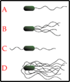
The different arrangements of bacterial flagella: A-Monotrichous; B-Lophotrichous; C-Amphitrichous; D-Peritrichous
-
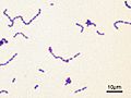
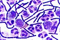
Streptococcus mutans visualised with a Gram stain.
-
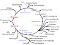
Phylogenetic tree showing the diversity of bacteria, compared to other organisms. Here bacteria are represented by three main supergroups: the CPR ultramicrobacterias, Terrabacteria and Gracilicutes according to recent genomic analyzes (2019).
-
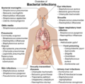
Overview of bacterial infections and main species involved.
-
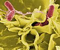
Colour-enhanced scanning electron micrograph showing Salmonella typhimurium (red) invading cultured human cells
-

Antonie van Leeuwenhoek, the first microbiologist and the first person to observe bacteria using a microscope.


._Natuurkundige_te_Delft_Rijksmuseum_SK-A-957.jpeg)
See also
 In Spanish: Bacteria para niños
