
Scanning electron microscope facts for kids


A scanning electron microscope (SEM) is a special type of electron microscope. It uses a focused beam of tiny particles called electrons. These electrons hit the surface of a solid object. When they hit, they create different signals. These signals give us lots of helpful information. We can learn about the object's shape, its tiny parts (atomic structure), and how well it conducts electricity.
When an electron touches the surface of an object, a few things can happen. It might bounce back (this is called backscattered). It could also be soaked up by the object. Or, it might be carried away by the object. When electrons are soaked up, they can make the atoms they hit a bit unstable. To become stable again, these atoms might let go of another electron. These new electrons are called secondary electrons. Or, the atoms might give off light to settle down.
Microscopes can have different tools called detectors. These detectors look for the different reactions that happen. The type of detector used depends on what information scientists want to find out about the object.
Contents
How Powerful Is an SEM?
The SEM can make things look incredibly big. How much it can magnify depends on how thin the electron beam can be. It can show details as small as 1 nanometer. That's about the size of 3 to 5 atoms! Imagine seeing something that tiny.
Magnets are used to control the electron beam. Other magnets help shape the beam. They also move it across the object being looked at. The SEM can magnify things from 30 times their actual size. It can go all the way up to 500,000 times bigger!
Why a Vacuum Is Needed
The electron beam is very sensitive. Air and water particles can mess it up. Because of this, the object being looked at must be placed in a vacuum. A vacuum is an empty space with no air.
The object also needs to be able to conduct electricity. This lets any electrons that aren't bounced back or soaked up move away. This means there are some limits on what kinds of objects can be studied. For example, if scientists want to look at plants or animals, they often have to coat them in gold first. This helps keep the sample from changing or breaking down during the scanning process.
Parts of an SEM
A scanning electron microscope has several important parts that work together:
The Electron Gun
This part is usually at the very top or bottom of the SEM. Its job is to shoot out a beam of electrons. These electrons are aimed at the object we want to examine.
The Lenses
The lenses in an SEM are not made of glass like in a regular microscope. Instead, they are made of strong magnets. These magnets bend and focus the electron beam. This helps create clear and detailed pictures of the object.
The Sample Chamber
This is the special area where the object you want to look at is placed. The SEM is very sensitive, so this chamber must be very strong. It needs to have no vibrations at all. This keeps the object perfectly still. If the object moves, the SEM can't make good pictures.
The Detector
Think of the detector as the "eyes" of the microscope. It's what helps create the super detailed pictures of the object's surface. There are many different types of detectors. Scientists choose the best one based on what information they want to get from the object.
The Vacuum Chamber
This chamber is very important. It keeps the electron beam from hitting air particles. If air particles were present, they would knock the electrons off course. This would make the pictures blurry and distorted.
Images for kids
-

Schottky-emitter electron source
-
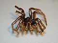
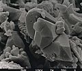
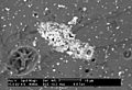
A spider sputter-coated in gold, having been prepared for viewing with an SEM
-

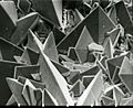
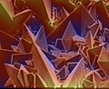
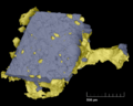
Low-temperature SEM magnification series for a snow crystal. The crystals are captured, stored, and sputter-coated with platinum at cryogenic temperatures for imaging.
-
Comparison of SEM techniques: Top: backscattered electron analysis – composition Bottom: secondary electron analysis – topography
-
Color cathodoluminescence overlay on SEM image of an InGaN polycrystal. The blue and green channels represent real colors, the red channel corresponds to UV emission.
-
Surface of a kidney stone
-
The same after re-processing of the color from the estimated topography
-
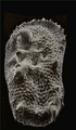
SEM image of a diagenetically altered discoaster
-
The same image after similar colorization
-
SEM image of Cobaea scandens pollen
-
The same after semi-automatic coloring. Arbitrary colors help identifying the various elements of the structure
-

Colored SEM image of Tradescantia pollen and stamens
-
Colored SEM image of native gold and arsenopyrite crystal intergrowth
-
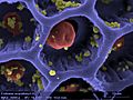
DDC-SEM of calcified particle in cardiac tissue - Signal 1 : SE
-
Signal 2 : BSE
-
Colorized image obtained from the two previous. Density-dependent color scanning electron micrograph SEM (DDC-SEM) of cardiovascular calcification, showing in orange a calcium phosphate spherical particle (denser material) and, in green, the extracellular matrix (less dense material)
-
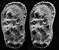
An SEM stereo pair of microfossils of less than 1 mm in size (Ostracoda) produced by tilting along the longitudinal axis.
-
From this pair of SEM images, the third dimension has been reconstructed by photogrammetry (using MountainsSEM software, see next image) ; then a series of 3D representations with different angles have been made and assembled into a GIF file to produce this animation.
-
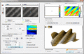
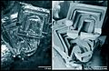
3D surface reconstruction of a (Ra = 3 µm) roughness calibration sample (as used to calibrate profilometers), from 2 scanning electron microscope images tilted by 15° (top left). The calculation of the 3D model (bottom right) takes about 1.5 second and the error on the Ra roughness value calculated is less than 0.5%.
-
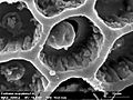
SEM image of a house fly compound eye surface at 450× magnification.
-
Detail of the previous image.
-
SEM 3D reconstruction from the previous using shape from shading algorithms.
-
Same as the previous, but with lighting homogenized before applying the shape from shading algorithms
-

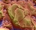
Colored SEM image of soybean cyst nematode and egg. The artificial coloring makes the image easier for non-specialists to view and understand the structures and surfaces revealed in micrographs.
-
Compound eye of Antarctic krill Euphausia superba. Arthropod eyes are a common subject in SEM micrographs due to the depth of focus that an SEM image can capture. Colored picture.
-
Ommatidia of Antarctic krill eye, a higher magnification of the krill's eye. SEMs cover a range from light microscopy up to the magnifications available with a Transmission electron microscopy (TEM). Colored picture.
-
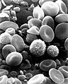
SEM image of normal circulating human blood. This is an older and noisy micrograph of a common subject for SEM micrographs: red blood cells.
-
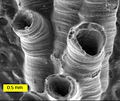
SEM image of a hederelloid from the Devonian of Michigan (largest tube diameter is 0.75 mm). The SEM is used extensively for capturing detailed images of micro and macro fossils.
-
Backscattered electron (BSE) image of an antimony-rich region in a fragment of ancient glass. Museums use SEMs for studying valuable artifacts in a nondestructive manner.
-
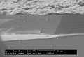
SEM image of the corrosion layer on the surface of an ancient glass fragment; note the laminar structure of the corrosion layer.
-
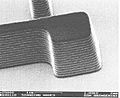
SEM image of a photoresist layer used in semiconductor manufacturing taken on a field emission SEM. These SEMs are important in the semiconductor industry for their high-resolution capabilities.
-
SEM image of the surface of a kidney stone showing tetragonal crystals of Weddellite (calcium oxalate dihydrate) emerging from the amorphous central part of the stone. Horizontal length of the picture represents 0.5 mm of the figured original.
-
Two images of the same depth hoar snow crystal, viewed through a light microscope (left) and as an SEM image (right). Note how the SEM image allows for clear perception of the fine structure details which are hard to fully make out in the light microscope image.
-
Epidermal cells from the inner surface of an onion flake. Beneath the shagreen-like cell walls one can see nuclei and small organelles floating in the cytoplasm. This BSE-image of a lanthanoid-stained sample was taken without prior fixation, nor dehydration, nor sputtering.


.gif)


See also
 In Spanish: Microscopio electrónico de barrido para niños
