
Brain facts for kids

The brain is an amazing organ that acts as the control center for the nervous system in almost all animals. It's made of special nervous tissue and is usually found in the head, close to important senses like sight, hearing, and smell. Think of it as your body's supercomputer!
Your brain is responsible for many incredible things. It takes in information from your senses, helps you think, understand, and learn new things. It also coordinates all your movements, from running and jumping to simply blinking your eyes. It even helps manage your body's hormones.
In humans, the outer part of the brain, called the cerebral cortex, has billions of tiny cells called neurons. Your cerebellum, which helps with coordination, has even more! Each neuron connects to thousands of others, sending signals through tiny fibers called axons and dendrites. These signals are like electrical messages, traveling super fast to different parts of your brain and body. The front part of your brain, the prefrontal cortex, is especially good at planning and making decisions.
Brains help animals respond quickly to changes around them. While simple actions like reflexes can happen in the spinal cord, complex behaviors and thinking need the brain to put all the information together. Scientists are still learning how millions of brain cells work together to create our thoughts and actions. They often compare the brain to a biological computer because it gathers, stores, and processes information from the world.
This article will explore the brains of different animals, especially vertebrates (animals with backbones). We'll look at how they are built, how they grow, and how they work. For more specific details about the human brain, you can check out that article.
How the Brain is Built

Brains come in many shapes and sizes across different animals. But they all share some basic building blocks. To study the brain, scientists often harden the soft tissue and slice it very thinly. This helps them see inside.
Inside the brain, you'll find areas of darker tissue called grey matter and lighter tissue called white matter. Grey matter is where most of the neuron cell bodies are, while white matter is mostly made of the long connecting fibers. Scientists also use special dyes to highlight different parts and see how they connect.
Brain Cells: Neurons and Glia

Your brain is made of two main types of cells: neurons and glial cells. Glial cells are like the brain's support team. They help neurons with structure, energy, and protection. Neurons are the stars of the show, though! They are the cells that send and receive messages.
Neurons are special because they can send signals over long distances. They have a long fiber called an axon that stretches out from the cell body. Axons can be very long; if a neuron's body were the size of a human, its axon could be over a kilometer long! These axons send fast electrical signals to other neurons.
Neurons talk to each other at tiny meeting points called synapses. When an electrical signal reaches a synapse, it releases a chemical called a neurotransmitter. This chemical then tells the next neuron what to do. Your brain has trillions of synapses, and they are always changing. This ability to change is how your brain learns and remembers things!
Many axons are covered in a fatty layer called myelin. Myelin acts like insulation on an electrical wire, making the signals travel much faster. Myelin is white, which is why areas of the brain with many myelinated axons are called white matter.
How Brains Evolved
Most animals, except for very simple ones like sponges, have a symmetrical body plan. This means they have a left and right side that are almost mirror images. Scientists believe all these animals came from a common ancestor that looked like a simple worm. This worm likely had a nerve cord running through its body with a larger cluster of nerves at the front, which was the first "brain."
Brains in Invertebrates
.jpg)
Invertebrates are animals without backbones, like insects, spiders, and octopuses. Their brains are very diverse. Some, like nematode worms, have simple brains. Others, like octopuses and squids, have surprisingly complex brains, the largest among invertebrates!
Scientists study the brains of certain invertebrates to learn more about how brains work. For example, fruit flies (Drosophila) are great for studying how genes affect brain development. The tiny worm Caenorhabditis elegans has a nervous system with exactly 302 neurons, always in the same places. This makes it perfect for mapping every single connection, like a complete wiring diagram!
Brains in Vertebrates

Vertebrates, animals with backbones, first appeared over 500 million years ago. Modern fish, amphibians, reptiles, birds, and mammals show how brains have grown in size and complexity over time. All vertebrate brains have the same basic parts, but some parts become much larger and more developed in mammals.
Generally, bigger animals tend to have bigger brains. But it's not a simple rule. Smaller animals often have proportionally larger brains compared to their body size. For example, primates (like monkeys and humans) have brains much larger than what you'd expect for their body size. Animals that hunt often have larger brains than their prey, which helps them plan hunting strategies.

All vertebrate brains start with a similar shape during early development. They begin as three swellings at the front of a tube, which become the forebrain, midbrain, and hindbrain. In fish and amphibians, these parts stay similar in size. But in mammals, the forebrain grows much larger, and the hindbrain develops a big part called the cerebellum.
Vertebrate brains are very soft and are protected by membranes called meninges and the skull. Special blood vessels form a blood–brain barrier that protects the brain from harmful substances.
Scientists divide the vertebrate brain into six main areas: the telencephalon (which includes the cerebral hemispheres), diencephalon (thalamus and hypothalamus), mesencephalon (midbrain), cerebellum, pons, and medulla oblongata. Some parts, like the cerebral cortex, are folded to fit more surface area into the skull.
Mammalian Brains
Mammalian brains are generally much larger than those of other vertebrates of the same body size. The biggest difference is in the forebrain, especially the cerebral cortex. In other vertebrates, the cortex is simpler. But in mammals, it becomes a complex, six-layered structure called the neocortex. Areas like the hippocampus and amygdala, important for memory and emotions, are also much more developed in mammals.
The corpus callosum is a thick band of nerves that connects the two halves of the brain in placental mammals, allowing them to communicate.
Primate Brains
| Species | EQ |
|---|---|
| Human | 7.4–7.8 |
| Common chimpanzee | 2.2–2.5 |
| Rhesus monkey | 2.1 |
| Bottlenose dolphin | 4.14 |
| Elephant | 1.13–2.36 |
| Dog | 1.2 |
| Horse | 0.9 |
| Rat | 0.4 |
The brains of humans and other primates are larger compared to their body size than most other mammals. Scientists use something called the encephalization quotient (EQ) to compare brain sizes fairly. Humans have a very high EQ, meaning our brains are much larger than expected for our body size.
Most of this extra size in primate brains comes from a huge expansion of the cerebral cortex, especially the prefrontal cortex. This part of the brain helps with planning, working memory, and making decisions. It's a much bigger part of the brain in primates, and especially in humans.
Brain Development

The brain grows in a very organized way, starting from a simple swelling in the early embryo. Neurons are made in special areas and then travel to their final spots. Once in place, their axons grow out, branching and extending until they reach their targets and form connections. Sometimes, too many neurons and connections are made at first, and then the unneeded ones are removed.
In vertebrates, the early steps of brain development are very similar across all species. A special strip of cells on the back of the embryo folds inward to form a hollow tube. This tube then swells at the front to create the forebrain, midbrain, and hindbrain. These parts then divide further, and new neurons and glial cells are generated in these areas.
Once a neuron is in its place, it sends out its dendrites and axon. The axon's tip, called a growth cone, senses its surroundings and is guided to the correct destination. Many genes help guide these axons to find their way.
The connections in your brain are not just set by your genes. Your experiences also play a huge role! At first, many connections are made, but then they are "pruned" or strengthened based on how much they are used. This process helps fine-tune the brain's wiring. For example, the signals from your eyes help create a precise map in your brain for vision.
In humans, most new neurons are made before birth. However, a few areas, like those involved in smell and memory, continue to make new neurons throughout life. Glial cells, on the other hand, are made throughout your entire life.
Both your genes and your experiences are important for how your brain develops. Genes set up the basic structure, but experiences help refine the connections, making your brain more complex. For example, animals raised in stimulating environments often have thicker cerebral cortices, meaning more connections, than those with less stimulation.
How the Brain Works
The brain's functions depend on neurons sending and receiving electrical and chemical signals. These signals are controlled by many processes, especially the interaction between neurotransmitters and receptors at synapses.
Chemical Messengers: Neurotransmitters
Neurotransmitters are chemicals released at synapses when an electrical signal arrives. They attach to special receptors on the next cell, changing its activity. Each neuron usually releases the same neurotransmitter at all its connections. Many medicines work by affecting these neurotransmitter systems.
The two most common neurotransmitters in the vertebrate brain are glutamate, which usually excites neurons, and gamma-aminobutyric acid (GABA), which usually calms them down. Because they are found almost everywhere, medicines that affect glutamate or GABA can have strong effects. For example, some medicines that help you sleep work by boosting GABA's effects.
Other neurotransmitters, like serotonin and dopamine, work in more specific areas of the brain and are involved in things like mood, arousal, and motivation.
Brain's Electrical Activity

As neurons send electrical signals, they create tiny electric fields. When many neurons work together, these fields can be strong enough to be measured from outside the head using tools like electroencephalography (EEG). EEG recordings show that your brain is always active, even when you're sleeping!
Brain activity changes depending on what you're doing. During deep sleep, the brain shows large, slow waves. When you're awake but relaxed, there are faster waves. When you're focused on a task, the activity looks more chaotic. Sometimes, if the brain's control mechanisms don't work right, electrical activity can become too high, leading to patterns seen during an epileptic seizure.
Brain's Energy Needs
Your brain uses a lot of energy! Even though it's only a small part of your body, it uses about 20-25% of your body's total energy in humans. Most of this energy goes into keeping neurons ready to send electrical signals. The brain mainly gets its energy from glucose (blood sugar) and oxygen. When you're thinking hard, active parts of your brain use a bit more energy, which is how brain imaging techniques like fMRI work.
What the Brain Does
The brain collects information from your senses. It uses this information to understand the world, combine it with your needs and memories, and then decide what actions to take. This complex process involves many different parts of the brain working together.
The brain's main job is to control an animal's actions in a coordinated way. It allows different muscles to work together and helps the body respond to things happening around it.
Sensing the World: Perception

Your brain receives information about light, sound, smells, tastes, temperature, and your body's position. Some animals have extra senses, like snakes that can sense heat or birds that can sense magnetic fields.
Each sense has special cells that detect information. For example, your eyes have cells that detect light. These cells send signals to your brain, which then processes them to create your experience of seeing, hearing, or smelling. Different parts of the brain specialize in processing different types of sensory information.
Controlling Movement
Motor systems are the parts of the brain that start body movements. Most voluntary muscles in your body are controlled by neurons in your spinal cord and hindbrain. These spinal neurons are controlled by signals coming down from your brain.
Your brain has several areas that send signals directly to the spinal cord. Lower brain areas control basic movements like walking or breathing. Higher areas, like the primary motor cortex in the front of your brain, allow for very precise and voluntary control of movements, like writing or playing an instrument. Other areas, like the cerebellum and basal ganglia, help refine and coordinate these movements.
| Area | Location | Function |
|---|---|---|
| Ventral horn | Spinal cord | Contains motor neurons that directly activate muscles |
| Oculomotor nuclei | Midbrain | Contains motor neurons that directly activate the eye muscles |
| Cerebellum | Hindbrain | Makes movements precise and well-timed |
| Basal ganglia | Forebrain | Helps choose actions based on what you want |
| Motor cortex | Frontal lobe | Directly activates spinal motor circuits for movement |
| Premotor cortex | Frontal lobe | Groups simple movements into coordinated patterns |
| Supplementary motor area | Frontal lobe | Organizes movements into sequences |
| Prefrontal cortex | Frontal lobe | Helps with planning and making decisions |
Sleep and Waking

Many animals have a daily cycle of sleeping and waking. A tiny part of the hypothalamus called the suprachiasmatic nucleus (SCN) acts as your body's main biological clock. It keeps time with a roughly 24-hour rhythm, influenced by light signals from your eyes.
Sleep involves big changes in brain activity. There are two main types of sleep: REM sleep (when you usually dream) and NREM sleep (usually without dreaming). During deep NREM sleep, your brain shows large, slow waves of activity.
Keeping Balance: Homeostasis
Your brain helps keep your body's internal conditions stable, like temperature, water content, and blood sugar levels. This process is called homeostasis. The hypothalamus plays a huge role in this. It receives information from sensors in your body and sends signals to correct any imbalances. It also controls the pituitary gland, which releases hormones to make changes throughout your body.
Motivation and Choices

Your brain's motivational system helps you seek things you need, like food, water, or shelter. It works using a reward-punishment system. When you do something that has good results, your brain's reward system is activated, making you more likely to repeat that behavior. If a behavior has bad results, your brain helps you avoid it in the future.
The basal ganglia, a group of structures at the base of the forebrain, are central to making these decisions. They control many motor systems and release inhibition when an action is chosen. The neurotransmitter dopamine is very important in the reward system.
Learning and Memory
Almost all animals can change their behavior based on experience. Scientists believe that learning and memory happen when the connections between neurons (synapses) change. This idea is called synaptic plasticity.
Neuroscientists recognize different types of learning and memory:
- Working memory is your brain's ability to hold temporary information, like remembering a phone number for a short time.
- Episodic memory is remembering specific events, like what you did last summer. The hippocampus is very important for forming these long-lasting memories.
- Semantic memory is remembering facts and relationships, like knowing that Paris is the capital of France. This is likely stored in the cerebral cortex.
- Instrumental learning is when rewards and punishments change your behavior. The basal ganglia are key here.
- Motor learning is improving your movements through practice, like learning to ride a bike. The cerebellum is especially important for this.
Brain Research

The study of the brain and nervous system is called Neuroscience. Psychology looks at the mind and behavior, while neurology treats brain diseases. Psychiatry focuses on mental health. Cognitive science combines neuroscience, psychology, computer science, and philosophy to understand the brain.
Scientists have studied the brain for a long time. The oldest brain ever found was in Armenia, over 5,000 years old, in the skull of a 12 to 14-year-old girl. It was well preserved due to the cave's climate.
Early thinkers wondered if the brain or heart was the center of thought. The ancient Greek physician Hippocrates believed the brain was the source of our joys, sorrows, and understanding. The Roman physician Galen showed that nerves connect the brain to all muscles.
A big step forward came with Luigi Galvani in the 1700s, who discovered that electricity could make a frog's leg move, showing nerves use electrical signals. Later, new ways to stain cells, like the Golgi stain, allowed scientists like Santiago Ramón y Cajal to see individual neurons and their complex shapes for the first time.

In the 20th century, advances in electronics helped scientists study the electrical properties of nerve cells. This led to understanding how neurons send signals. The invention of computers also helped scientists think of the brain as an information-processing system.
Today, scientists use many tools to study the brain. They can record the electrical activity of many brain cells at once, use genetic engineering to change specific genes, and use neuroimaging techniques like fMRI to see which parts of the human brain are active during different tasks. They also study what happens when specific brain areas are damaged, which helps them understand what those areas do.
The Human Brain Project is a large research effort that aims to create a detailed computer model of the entire human brain. This is a huge challenge, but it could help us understand the brain like never before.
Brain diseases
Brain diseases are illnesses that affect the brain and how it works.
Some brain diseases, like Alzheimer's disease, can cause people to slowly lose their memories and have trouble thinking clearly. It's as if the brain's filing system for memories starts to get mixed up. Other diseases, like Parkinson's, can make a person's hands shake or make their movements stiff and slow because the brain has trouble sending the right signals to the muscles. There are also diseases like epilepsy, which can cause sudden "electrical storms" in the brain called seizures. These can make a person's body jerk or cause them to stare blankly for a few moments.
When the brain gets inflamed, the immune system gets confused and starts attacking the brain’s own healthy cells. This can happen because of an infection, like meningitis, or sometimes because the body’s defenses make a mistake. This inflammation can lead to different diseases. For example, it can cause encephalitis, which is like having a bad flu that affects the brain, leading to headaches, confusion, or even seizures. Inflammation is also a key part of multiple sclerosis (MS), where it damages the protective coating around nerve fibers, slowing down messages between the brain and the body. Scientists are also learning that inflammation might play a role in other brain conditions, showing just how important it is to keep our body’s defense system in balance.
The good news is that doctors and scientists are working hard to understand these diseases. While not all brain diseases can be cured yet, many can be treated with medicine, therapy, or healthy lifestyle choices. Things like eating nutritious food, getting enough sleep, and protecting your head by wearing a helmet when you ride your bike are all great ways to help keep your brain healthy for a long time.
Related pages
Images for kids
-
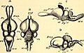
Anatomical comparison between the brain of a lizard (A and C) and the brain of a turkey (B and D).
-
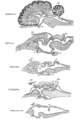
Comparison of Vertebrate Brains: Mammalian, Reptilian, Amphibian, Teleost, and Ammocoetes.
-
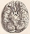
Andreas Vesalius' Fabrica, published in 1543, showing the base of the human brain.
_(20482913440).jpg)


See also
 In Spanish: Cerebro para niños
