
Bone facts for kids

Bones are the strong, stiff parts that make up the skeleton in humans and animals with backbones. They are like the frame of your body. Without bones, we couldn't stand up or move around. Bones also protect the soft organs inside us. For example, your skull protects your brain, and your rib cage protects your heart and lungs. Your jaw and cheekbones help support your face muscles, which let you smile! Did you know that a human's neck has the same number of bones as a giraffe's?
Contents
What Bones Do
Bones are the main framework of your body. Imagine trying to move without them – you'd just be a pile of soft parts on the ground! Bones are super important for keeping you upright and letting you move. They also protect your insides. Your rib cage keeps your heart and lungs safe. Your skull protects your brain. Your pelvis (hip bones) protects your reproductive organs. And your vertebrae (backbones) protect your spinal cord.
To keep your bones strong, you need to exercise regularly. Eating foods rich in calcium also helps a lot. Think of milk and dark leafy greens like spinach! Inside your bigger bones, there's something called red bone marrow. This amazing stuff makes the red blood cells your body needs to carry oxygen.
How Bones Are Built
Quick facts for kids Osteon |
|
|---|---|
 |
|
| Diagram of compact bone from a transverse section of a long bone's cortex. | |
| Latin | Osteon |
| Gray's | subject #18 89 |
Bones aren't just solid all the way through. They have a flexible part called a matrix and hard minerals. The matrix is mostly made of stretchy collagen fibers.
The tough outer layer of bones is called cortical bone or compact bone. It's what makes bones look smooth, white, and solid. About 80% of an adult's bone mass is compact bone. This strong layer helps support your whole body. It also protects your organs and acts as levers for your muscles to move. Compact bone stores and releases important chemicals, especially calcium. It's made of tiny columns called osteons. Each column has layers of special bone cells. Some cells, called osteoblasts, help build new bone. Others, like osteocytes and osteoclasts, help reshape and break down old bone.
Inside, you'll find cancellous bone or spongy bone. It looks like a sponge with many small holes. Spongy bone is usually found at the ends of long bones, near joints, and inside your backbones. It often contains red bone marrow. This marrow creates blood cells for your blood system. It also makes lymphocytes for your immune system, which fights off sickness.
Types of Bones

Your body has five main types of bones: long, short, flat, irregular, and sesamoid.
- Long bones are much longer than they are wide. They have a main shaft and rounded ends. Most of your arm and leg bones are long bones. This includes the bones in your fingers and toes. They are mostly made of compact bone, with spongy bone at the ends.
- Short bones are roughly cube-shaped. They have a thin layer of compact bone around a spongy inside. Short bones give you stability and support, plus a little bit of movement. The bones in your wrist and ankle are short bones.
- Flat bones are thin and usually curved. They have two layers of compact bone with a spongy layer in between. Most of your skull bones are flat bones. Your sternum (breastbone) is also a flat bone.
- Sesamoid bones are small bones found inside tendons. They help increase the power of muscles by changing the angle of the tendon. Your patella (kneecap) is a good example of a sesamoid bone.
- Irregular bones don't fit into the other categories. They have complex and unusual shapes. They also have thin layers of compact bone around a spongy inside. Bones in your spine, pelvis, and some skull bones are irregular bones.

How Bones Grow

.jpg)
The process of bone forming is called ossification. This happens in two main ways when a baby is developing before birth:
- Intramembranous ossification is when bone forms directly from connective tissue. This is how most of the flat bones in your skull, and your jawbones, are made.
- Endochondral ossification is when bone forms from cartilage. This is how your long bones and most other bones in your body grow. Cartilage acts like a model that slowly turns into bone.
Bone Breaks (Fractures)

A fracture is simply a broken bone. Bones can break if a lot of force is applied to them. They can also break from repeated stress over time. Sometimes, bones can break more easily if they are weak. This can happen with conditions like osteoporosis, which makes bones brittle.
Common places for bones to break include the wrist and hip. Breaks in the backbones (vertebrae) can happen from strong impacts. Not all fractures are painful right away. If a bone breaks through the skin, it's called an open fracture. Sometimes, if a piece of bone is missing, doctors can use bone grafting to replace it.
Doctors use special tools to check for broken bones. These include X-rays, CT scans, and MRI scans.
Cool Facts About Bones
- When you are born, you have about 300 bones! As you grow, many of these bones join together. By the time you're an adult, you have 206 separate bones.
- The Greek word for bone is "osteon." That's why you see "osteo" in many bone-related words, like osteopathy.
- The biggest bone in your body is the femur, or thigh-bone. The smallest bone is the stapes, found in your middle ear. It's tiny, like a grain of rice!
- Some animals, especially plant-eaters, sometimes eat bones. This is called osteophagy. They do this to get important minerals like phosphate that they might be missing.
Images for kids
-
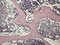
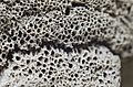
Micrograph of cancellous bone
-
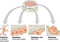
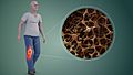
Bone cells
-

Skeletal System of Human Body
-
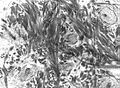
Transmission electron micrograph of decalcified woven bone matrix displaying characteristic irregular orientation of collagen fibers
-
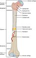
Structure of a long bone
-
Reduced bone mineral density in Osteoporosis (R), increasing the likelihood of fractures
-
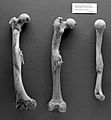
Human femurs and humerus from Roman period, with evidence of healed fractures
-
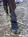
Skeletal fluorosis in a cow's leg, due to industrial contamination
-
Leg and pelvic girdle bones of bird
-
Structure detail of an animal bone


See also
 In Spanish: Hueso para niños
