
Heart facts for kids
Quick facts for kids Heart |
|
|---|---|
 |
|
| An illustration of the front view of the human heart | |
| Latin | cor |
| System | Circulatory |
| Artery | Aorta, pulmonary trunk and right and left pulmonary arteries, right coronary artery, left main coronary artery |
| Vein | Superior vena cava, inferior vena cava, right and left pulmonary veins, great cardiac vein, middle cardiac vein, small cardiac vein, anterior cardiac veins |
| Nerve | Accelerans nerve, vagus nerve |
The heart is a powerful muscle in humans and other animals that works like a pump. Its main job is to push blood through tubes called blood vessels to every part of your body. This network of the heart and blood vessels is called the circulatory system. The blood it pumps carries oxygen and nutrients to all your cells. It also picks up waste products, like carbon dioxide, to be removed from the body.
In humans, the heart is about the size of a fist. It is located in the middle of the chest, between the lungs. The heart is divided into four main sections, called chambers. Heart valves act like one-way doors between these chambers to keep blood flowing in the right direction. The heart is protected by a sac called the pericardium.
The heart's beat is controlled by a natural pacemaker that sends out electrical signals. This makes the heart muscle squeeze (contract) in a regular rhythm. A normal resting heart rate for an adult is about 72 beats per minute. Exercise makes the heart beat faster to supply the body with more oxygen, which is very good for heart health.
How the Heart is Built


Location and Shape
The human heart is found in the center of the chest, in a space called the mediastinum. It sits behind the breastbone (sternum) and between the two lungs. The back of the heart is near the spine.
The heart is shaped like a cone, with the wide part at the top and a pointy tip, called the apex, at the bottom. The apex points toward the left side of the chest. This is why you usually feel your heartbeat on the left side. An adult heart weighs about 250–350 grams (9–12 oz).
The Four Chambers
The heart is divided into four hollow areas called chambers. There are two chambers on the top and two on the bottom.
- The atria (singular: atrium) are the two upper chambers. They are the receiving rooms for blood coming back to the heart. The right atrium receives blood from the body, and the left atrium receives blood from the lungs.
- The ventricles are the two lower chambers. They are the pumping rooms that send blood out of the heart. The right ventricle pumps blood to the lungs, and the left ventricle pumps blood to the rest of the body.
The left ventricle's wall is much thicker and stronger than the right ventricle's. This is because it has to pump blood to the entire body, while the right ventricle only pumps blood to the nearby lungs. A wall of muscle called the septum separates the left and right sides of the heart.
The Heart's Valves

.svg)
The heart has four valves that act like one-way gates. They open to let blood move forward and then snap shut to prevent it from flowing backward. This ensures that blood always moves in one direction.
- The tricuspid valve is between the right atrium and the right ventricle.
- The pulmonary valve is between the right ventricle and the pulmonary artery, which leads to the lungs.
- The mitral valve is between the left atrium and the left ventricle.
- The aortic valve is between the left ventricle and the aorta, the body's main artery.
Tiny, strong cords called chordae tendineae are attached to the tricuspid and mitral valves. They act like anchors to stop the valves from being pushed back too far when the ventricles pump.
The Heart Wall

The wall of the heart is made of three layers:
- The endocardium is the smooth, inner lining of the heart chambers and valves.
- The myocardium is the thick, muscular middle layer. This is the part of the heart that contracts to pump blood. The muscle cells are arranged in a special swirling pattern that helps the heart pump very effectively.
- The epicardium is the thin, outer layer of the heart wall.
Surrounding the heart is a protective, double-layered sac called the pericardium. It contains a small amount of fluid that allows the heart to beat smoothly without rubbing against other organs.
Blood Supply for the Heart

Just like any other muscle in the body, the heart needs its own supply of oxygen-rich blood to work. The blood vessels that do this job are called the coronary arteries. These arteries branch off from the aorta and spread over the surface of the heart, delivering the oxygen and nutrients the heart muscle needs to keep pumping.
The Heart's Big Job: Pumping Blood
The heart's main function is to pump blood around the body. This process happens in two main circuits that are connected. This is called a double circulatory system.
The Journey of Blood

Imagine you are a drop of blood starting your journey.
- To the Lungs: Oxygen-poor blood (shown in blue in many diagrams) returns from the body and enters the right atrium. The heart pumps this blood down into the right ventricle. The right ventricle then pumps the blood out to the lungs.
- In the Lungs: In the lungs, the blood drops off carbon dioxide and picks up fresh oxygen.
- Back to the Heart: This new, oxygen-rich blood (shown in red) travels from the lungs back to the heart, entering the left atrium.
- To the Body: The heart pumps the blood down into the powerful left ventricle. The left ventricle then pumps this oxygen-rich blood into the aorta, the body's largest artery. From the aorta, the blood travels to the rest of the body to deliver oxygen to all your cells.
After delivering oxygen, the blood becomes oxygen-poor again and travels back to the right atrium to start the journey all over. This whole cycle happens with every single heartbeat.
The Heartbeat

A single heartbeat is made up of two main phases. This is called the cardiac cycle.
- Systole is the squeezing phase. The ventricles contract, pushing blood out to the lungs and the rest of the body.
- Diastole is the relaxing phase. The ventricles relax and fill up with blood from the atria.
When you listen to a heart with a stethoscope, you hear a "lub-dub" sound. The "lub" sound (S1) is made by the tricuspid and mitral valves closing at the start of systole. The "dub" sound (S2) is made by the aortic and pulmonary valves closing at the start of diastole.
The Heart's Electrical Spark

The heart has its own electrical system that tells it when to beat. The "spark" starts in a special group of cells called the sinoatrial node (SA node), also known as the heart's natural pacemaker. The SA node is located in the right atrium.
It sends out a regular electrical signal that spreads across the atria, causing them to contract. The signal then travels to the ventricles, telling them to contract a moment later. This coordinated signal ensures that the heart beats in a steady, even rhythm.
Heart Rate
Your heart rate is the number of times your heart beats per minute (bpm). A typical resting heart rate for an adult is between 60 and 100 bpm. Children and teenagers often have faster heart rates.
Many things can affect your heart rate. When you exercise, your heart beats faster to pump more oxygen to your muscles. When you are resting or sleeping, it slows down. Feelings like excitement or fear can also make your heart beat faster.
Keeping Your Heart Healthy
Keeping your heart healthy is one of the most important things you can do for your body. A healthy heart allows you to run, play, and live an active life. Doctors who specialize in the heart are called cardiologists.
There are several simple things you can do to take care of your heart:
- Eat a balanced diet: Eating plenty of fruits, vegetables, and whole grains is good for your heart.
- Stay active: Regular exercise, like running, swimming, or playing sports, makes your heart muscle stronger.
- Avoid smoking: Smoking is very harmful to the heart and blood vessels.
- Get enough sleep: Rest is important for your overall health, including your heart.
Doctors can check on your heart's health using tools like a stethoscope to listen to your heart sounds and an ECG to record its electrical activity.
Discovering the Heart's Secrets

For thousands of years, people knew the heart was important, but they didn't fully understand how it worked. Ancient Egyptians believed the heart was the center of thought and emotion.
A major breakthrough came in 1628 when an English doctor named William Harvey correctly described how blood circulates through the body. He showed that the heart acts as a pump to push blood in a continuous loop.
In 1967, a South African surgeon named Christiaan Barnard performed the first successful human-to-human heart transplant. This was a major event in medical history and opened the door for new ways to treat serious heart problems.
The Heart as a Symbol

Besides being a vital organ, the heart is also a powerful symbol. For centuries, people have connected the heart with feelings of love, courage, and emotion. The well-known heart shape is used all over the world, especially on Valentine's Day, to represent love.
When we talk about a "broken heart," we are using the heart as a symbol for feelings of sadness and loss. The heart is a central symbol in art, poetry, and music.
Hearts in the Animal Kingdom
Humans and other mammals have a four-chambered heart. But in the animal kingdom, hearts come in different shapes and sizes.
- Fish have a simpler, two-chambered heart. It has one atrium and one ventricle. It pumps blood to the gills to get oxygen and then out to the rest of the body.
- Amphibians (like frogs) and most reptiles (like lizards) have a three-chambered heart with two atria and one ventricle. There is some mixing of oxygen-rich and oxygen-poor blood in the single ventricle.
- Crocodiles are an exception among reptiles; they have a four-chambered heart, much like birds and mammals.
- Insects have a very different system. Their "heart" is a long tube that pumps a fluid called haemolymph around their body in an open circulatory system.
Images for kids
-
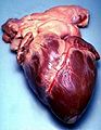
A look inside the human heart.
-
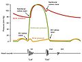
This graph shows the pressure changes in the heart during one heartbeat.
-
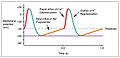
An electrical signal from the heart's pacemaker starts each beat.
-

A stethoscope is used to listen to the sounds of the heart.
-
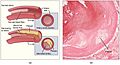
A buildup of plaque can narrow arteries, making it harder for blood to flow.
-
An ultrasound image showing the heart's valves in action.
-
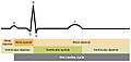
The ECG records the electrical activity that causes the heart to beat.


See also
 In Spanish: Corazón para niños
