
Inferior vena cava facts for kids
Quick facts for kids Inferior vena cava |
|
|---|---|
[[Image:
 |
|
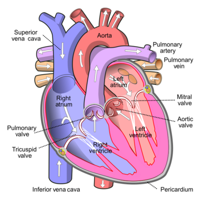
| Anterior (frontal) view of the opened heart. White arrows indicate valid blood flow. | |
 |
|
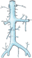
| Superior vena cava, inferior vena cava, azygos vein and their tributaries | |
| Latin | vena cava inferior |
| Artery | Abdominal aorta |
The inferior vena cava (IVC) is a very large vein in your body. It acts like a major highway for blood, carrying blood that has been used by your lower body and middle body back to your heart. This blood is "deoxygenated," meaning it has given up its oxygen to your body's cells. The IVC delivers this blood directly to the right atrium, which is one of the heart's upper chambers.
The IVC is formed when two other large veins, the right and left common iliac veins, join together. This usually happens around the level of your fifth lumbar vertebra, which is in your lower back. It's called "inferior" because it carries blood from the lower part of your body, unlike the superior vena cava which handles blood from your upper body. Together, these two venae cavae are the main veins that bring deoxygenated blood back to your heart.
The inferior vena cava is a large vein located behind your abdominal cavity (belly area). It runs along the right side of your vertebral column (backbone). It enters the lower right, back side of your heart. The name "vena cava" comes from Latin words: vena means "vein" and cavus means "hollow."
Contents
How the IVC is Built
The IVC starts when the left and right common iliac veins connect. It collects blood and brings it to the right atrium of your heart. Other veins also join the IVC, like the azygos vein (which runs along the right side of your backbone) and vein networks near your spinal cord.
The inferior vena cava begins behind your abdomen at about the level of your L5 lumbar vertebra. It then passes through a special opening in your thoracic diaphragm (the muscle that helps you breathe) at the level of your T8 or T9 thoracic vertebra. It runs to the right of your descending aorta, which is a major artery.
Veins that Join the IVC
Many smaller veins, called tributaries, drain their blood into the inferior vena cava. Here are some of the main ones:
- The hepatic veins and inferior phrenic vein join the IVC near your chest (T8 level).
- The right suprarenal vein (from your adrenal gland) and renal veins (from your kidneys) join around your lower back (L1 level).
- The right gonadal vein (from your reproductive organs) joins around L2.
- Several lumbar veins (from your lower back) join along its path (L1-L5).
- Finally, the common iliac veins from your legs join at the very bottom (L5).
Because the IVC is on the right side of your body, some veins join it directly on the right side. On the left side, some veins, like the left gonadal and suprarenal veins, first drain into the left renal vein, which then drains into the IVC.
How the IVC Develops
When a baby is developing inside its mother, the inferior vena cava and the right atrium are separated by a small flap called the Eustachian valve. In adults, this valve usually disappears completely or remains as a very tiny fold.
Different Ways the IVC Can Form
Sometimes, the way the IVC forms can be a bit different from person to person. About 8.7% of people have some variations in their IVC. These differences happen during development, usually between the 4th and 8th weeks of pregnancy.
The IVC is made up of four main parts that come from different blood vessels joining together. For example, in about 0.2% to 3% of people, there might be a "duplicated IVC," meaning there are two main IVCs below the kidney veins instead of just one. Most of the time, these variations don't cause any problems. However, doctors need to know about them if they are planning complex surgeries to avoid issues. Doctors can use special scans like ultrasound, CT, or MRI to see these variations.
What the IVC Does
The inferior vena cava is a vein. Its main job is to carry deoxygenated blood from the lower half of your body back to the right atrium of your heart. This blood is then pumped to your lungs to pick up oxygen.
The superior vena cava does the same job but for the upper half of your body.
Health Issues with the IVC
Problems with the IVC usually happen when something presses on it, rather than it breaking (which is very rare). Since the IVC has low pressure inside, it can be easily squeezed.
Things that can press on the IVC include:
- A very large aorta (a major artery).
- A pregnant uterus (this is why pregnant women who are unconscious are often turned onto their left side, to take pressure off the IVC and help blood flow back to the heart).
- Growths in the belly area, like certain types of cancer.
Sometimes, if blood flow through the IVC is restricted, it can even cause someone to faint.
It's rare for the inferior vena cava to get blocked, but if it does, it's a serious medical emergency. This can be linked to deep vein thrombosis (blood clots in deep veins), special filters placed in the IVC, or certain medical procedures.
If the vena cava is injured, for example, from trauma, it can lead to very serious blood loss and is often fatal.
Images for kids
-
Inferior vena cava
-
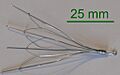
Image of an inferior vena cava filter
-
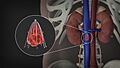
Image showing an inferior vena cava filter in its position


See Also
- Atriocaval shunt
- Inferior vena cava syndrome
- Recovery position
