
Cartilage facts for kids
Quick facts for kids Cartilage |
|
|---|---|
 |
|
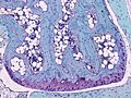
| Hyaline cartilage showing chondrocytes and organelles, lacunae and matrix |
Cartilage is a special kind of flexible tissue found in your body and in other animals. It's not as hard as bone but it's tougher than muscle. You can find cartilage in many places, like in your ear, nose, and between your bones in joints like your elbows, knees, and ankles. It's also in your rib cage and the discs between your vertebrae in your spine.
Cartilage is made of special cells called chondroblasts. These cells create a lot of surrounding material called the extracellular matrix. This matrix is made of strong fibers (like collagen and elastin) and a jelly-like substance. When chondroblasts get surrounded by this matrix, they become chondrocytes. These chondrocytes live in tiny spaces called lacunae.
Unlike most other tissues in your body, cartilage does not have blood vessels. This means it doesn't get a direct supply of blood. Because of this, cartilage heals very slowly if it gets damaged. The chondrocytes get their nutrients by absorbing them from nearby fluids, especially when the cartilage is squeezed or moved.
Contents
How Cartilage Grows
When a baby is developing inside its mother (this is called embryogenesis), its skeletal system starts as cartilage. This process, called chondrification or chondrogenesis, is how cartilage forms from early body tissue. The special cells (chondroblasts) begin to make the extracellular matrix, forming the cartilage.
Looking at Cartilage Inside the Body
It's tricky to see cartilage clearly with regular x-rays because it doesn't absorb them well. Doctors sometimes inject a special dye into a joint. This dye helps the cartilage show up better on the X-ray.
For a clearer view, doctors often use MRI (Magnetic Resonance Imaging). MRI is a very good way to see cartilage because it can show its shape, size, and even its chemical makeup. This is important for finding problems like injuries or diseases in joints.

Doctors use MRI to check cartilage for many reasons. For example, they look for Osteoarthritis, which is when cartilage wears away. They also check for injuries, inflammation, or other conditions that affect the joints. Newer MRI machines with stronger magnets can create even better and more detailed images of cartilage. This helps doctors make more accurate diagnoses, especially for smaller joints.
Types of Cartilage
There are three main types of cartilage, and they each have slightly different amounts of fibers and substances in their matrix:
- Elastic cartilage: This type is very flexible because it has many elastic fibers. You can find it in your outer ear and parts of your larynx (voice box).
- Hyaline cartilage: This is the most common type. It's strong but also flexible. It covers the ends of bones in your joints, helps form your nose, and is in your trachea (windpipe).
- Fibrocartilage: This is the strongest type of cartilage, with lots of tough collagen fibers. It's found in places that need to withstand a lot of pressure, like the discs between your vertebrae in your spine and in your knees.

Cartilage Problems and Treatments
Several conditions can affect cartilage. Here are some common ones:
- Osteoarthritis: This happens when the cartilage covering the ends of bones in a joint wears down. It can cause pain and make it hard to move the joint. It's often called "wear and tear" and usually affects joints that are used a lot. Sometimes, a severely damaged joint might need to be replaced with an artificial one in a surgery called Arthroplasty.
- Traumatic injury: Cartilage in the knee is often injured, especially during sports. Doctors are working on ways to repair or replace damaged knee cartilage.
- Achondroplasia: This is a genetic condition where cartilage in the long bones doesn't grow properly during childhood. It leads to dwarfism.
- Costochondritis: This is when the cartilage connecting your ribs to your breastbone gets inflamed, causing chest pain.
- Spinal disc herniation: The discs between your vertebrae have a soft center surrounded by tough cartilage. If this disc ruptures, the soft part can push out and press on nearby nerves, causing back pain.
- Relapsing polychondritis: This is a rare condition where the body's immune system attacks and destroys cartilage, especially in the nose and ears.
Sometimes, growths or tumors can form in cartilage tissue. These can be harmless (called chondroma) or cancerous (called chondrosarcoma).
One interesting thing about cartilage is that it acts like a barrier. It can prevent certain immune cells from entering it. This means that cartilage can sometimes be moved from one person to another without the body rejecting it, which is important for transplantation.
Cartilage Repair
Cartilage has a hard time repairing itself. Because its cells (chondrocytes) are stuck in their tiny spaces and it has no blood supply, new cartilage grows very slowly. Often, damaged hyaline cartilage is replaced by a tougher, less flexible type called fibrocartilage scar tissue.
However, scientists and doctors are working on new ways to help cartilage heal. They are developing techniques like bioengineering to grow new cartilage in labs. This involves using a special framework and cultured cells to create artificial cartilage that could one day replace damaged tissue.
Cartilage in Animals
Cartilaginous fish
Some fish, like sharks, rays, and skates, have skeletons made entirely of cartilage instead of bone! These are called cartilaginous fish.
Invertebrate cartilage
Even some animals without backbones, called invertebrates, have cartilage tissue. You can find it in creatures like horseshoe crabs, marine snails, and cephalopods (like octopuses and squids).
Images for kids
-
Section from mouse joint showing cartilage (purple)
-

Human skeleton with articular cartilage shown in blue


See also
 In Spanish: Tejido cartilaginoso para niños
