
Osteocyte facts for kids
Quick facts for kids Osteocyte |
|
|---|---|
 |
|
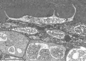
| Transverse section of a bone | |
 |
|
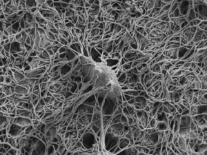
| Illustration showing a single osteocyte | |
| Latin | osteocytus |
An osteocyte is a special type of bone cell. It's shaped a bit like a flattened oval with many tiny "arms" or branches. These cells are the most common type found in grown-up bones. They can live for a very long time, sometimes as long as the person or animal they are part of! Your body has about 42 billion of these amazing cells. Osteocytes don't divide into new cells, and they usually live for about 25 years. They start out as other bone cells called osteoblasts, which then change into osteocytes.
Osteocytes and their "arms" live inside tiny spaces within the bone. The main part of the cell sits in a space called a lacuna (which means "pit" in Latin). Their long arms reach out through even tinier tunnels called canaliculi. Think of osteocytes as osteoblasts that got "trapped" inside the bone material they helped create. They stay connected to each other through their long arms, forming a network. This network helps them share nutrients and get rid of waste.
Even though osteocytes don't make much new bone material and can't divide, they are very important for keeping your bones healthy. They help with the daily repair and renewal of bone. They can even break down small amounts of bone when needed, in a process called osteocytic osteolysis.
What do osteocytes look like?
Osteocytes have a unique star-like shape. They are about 7 micrometers deep and wide, and about 15 micrometers long. (A micrometer is super tiny – a millionth of a meter!) The main body of the cell can be between 5 and 20 micrometers across. Each cell has many "arms" or processes, usually 40 to 60 of them. These arms help the cells connect to others, usually about 20 to 30 micrometers away.
A mature osteocyte has one main control center, called a nucleus. This nucleus usually has one or two smaller parts inside it called nucleoli. The cell also has smaller versions of other cell parts, like the endoplasmic reticulum, Golgi apparatus, and mitochondria. These parts help the cell do its job, even though they are smaller than in some other cells.
The "arms" of the osteocyte spread out like spokes on a wheel. They reach towards the surfaces of the bone or towards special canals that carry blood vessels. Together, the cell bodies in their lacunae and the arms in their canaliculi form a huge network. This network is spread throughout the hard, mineralized bone material.
How do osteocytes develop?
Scientists have found osteocytes in the bones of ancient jawless fish that lived millions of years ago! This shows that these cells have been around for a very long time.
When new bone is being made, a cell called an osteoblast helps create the bone material. As it works, the osteoblast can get surrounded and "buried" by the new bone material. When this happens, it changes and becomes an osteocyte. Even when it's buried, it keeps its connections to other osteoblasts and new osteocytes through its long arms.
The exact way an osteoblast turns into an osteocyte is still being studied. However, we know that certain molecules and conditions are important for this change. For example, about 10-20% of osteoblasts will turn into osteocytes. The osteoblasts that are going to become osteocytes slow down their bone-making work. Then, their neighbors, who are still working hard, bury them in the new bone material.
This change from a moving osteoblast to a trapped osteocyte takes about three days. During this time, the cell makes a lot of bone material – about three times its own size! As it becomes an osteocyte, the cell body actually shrinks by about 70% compared to its original osteoblast size. It also changes its shape from a roundish cell to a star-shaped cell with many arms reaching out.
Osteocytes seem to be good at surviving with less oxygen. This makes sense because they are buried deep inside the bone, where oxygen supply might be limited. The amount of oxygen available might even affect how osteoblasts turn into osteocytes.
What do osteocytes do?
Even though osteocytes are relatively quiet cells, they are very active in other ways. They can create and change molecules, and they can send signals over long distances, similar to how your nervous system works! They are the most common cell type in bone. For example, in cow bones, there are about 31,900 osteocytes in every cubic millimeter!
Osteocytes are super important for controlling how much bone you have. They act like sensors. If a bone breaks, osteocytes can produce signals that help with healing. When scientists have removed osteocytes in experiments, bones showed more breakdown, less new bone formation, and became weaker. This shows how vital osteocytes are for bone health.
These cells are like the "mechanosensors" of your bones. This means they can sense physical forces and stress on the bone. When you move or put weight on your bones, osteocytes feel it. They then send signals to other bone cells, like osteoblasts (which build bone) and osteoclasts (which break down bone). This helps control where and when bone needs to be built up or broken down. For example, osteocytes can send a signal that tells osteoblasts to start building new bone.
Osteocytes also play a key role in managing important minerals in your body, like phosphates. They produce special proteins, such as sclerostin, which help control how these minerals are used in your bones.
See also
- List of human cell types derived from the germ layers
- List of distinct cell types in the adult human body
