
Image: Parasite140015-fig2 Protoopalina pingi (Opalinidae) Microscopy
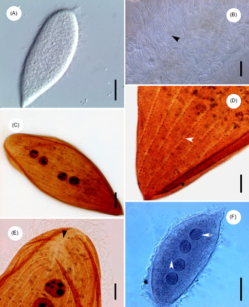
Description: Protoopalina pingi Nie, 1935 (Opalinidae). Figure 2 of paper. Light microscope images of Protoopalina pingi Nie, 1935. (A) Living specimens, showing the normal trophozoites of P. pingi. Scale bar = 20 μm. (B) Living specimens, showing the flagella covering the body (arrowhead). Scale bar = 5 μm. (C) Specimens stained with Protargol, showing the somatic kineties and the nuclei with distributed nucleoli. Scale bar = 10 μm. (D) Specimens stained with Protargol, showing the somatic kineties in the posterior extremity (arrowhead). Scale bar = 5 μm. (E) Specimens stained with Protargol, showing the falx region in the anterior extremity (arrowhead). Scale bar = 5 μm. (F) Specimens stained with Heidenhain’s haematoxylin, showing the nuclei (arrow) and the corpuscles of uneven size (arrowhead). Scale bar = 20 μm.
Title: Parasite140015-fig2 Protoopalina pingi (Opalinidae) Microscopy
Credit: Li, W., Wang, C., Huang, F., Li, M., Nilsen, F., Liu, H. & Xu, J. 2014: Redescription of Protoopalina pingi Nie, 1935 inhabiting the recta of Hylarana guentheri and Pelophylax nigromaculatus in China. Parasite, 21, 46. doi:10.1051/parasite/2014021
Author: Weidong Li, Chong Wang, Feng Huang, Ming Li, Frank Nilsen, Huiyu Liu and Jianlong Xu
Usage Terms: Creative Commons Attribution 4.0
License: CC BY 4.0
License Link: https://creativecommons.org/licenses/by/4.0
Attribution Required?: Yes
Image usage
The following page links to this image:
