
Image: Pezophaps limb bones

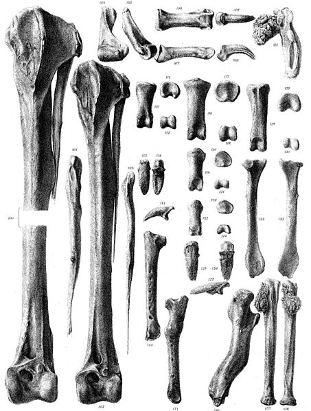
Description: Fig. 100. Left tibia, with fibula attached, from front (c??). Fig. 101. The same fibula, from behind. Fig. 102. Left tibia, with fibula attached, from front (??). Fig*. 103. The same fibula, from behind. Figs. 104, 105. Eight posterior metatarsus, from behind and outside (cH). Figs. 106, 107. Proximal phalanx of right hallux, from above and side (cH). Figs. 108, 109. Ungual phalanx of right hallux, from above and side (^i). Figs. 110-112. Proximal phalanx of right outer toe, from above and from each end ( cH). Figs. 113-115. Ungual phalanx of right outer toe, from above, below, and side (cH). Figs. 116-118. Proximal phalanx of right middle toe, from above and from each end ( 6 %). Figs. 119-121. Second phalanx of right middle toe, from above and from each end ( 6%). Figs. 122-124. Third phalanx of right middle toe, from above and from each end (tf?). Figs. 125-127. Ungual phalanx of right middle toe, from above, below, and side (tf?). Figs. 128-130. Proximal phalanx of right inner toe, from above and from each end ( 6 ?). Fig. 131. Eight metacarpal, lower view (<$V). (From a second specimen.) Figs. 132, 133. Left scapula, outer and inner surface (21). Fig. 134. Eight ulna, upper view, showing impressions of secondary quill-feathers (S*?). Fig. 135. Left ulna, upper view, showing the same, and also mark of fracture and healing (c?1). Fig. 136. Eight coracoid, side view, showing mark of fracture and healing (cH). Figs. 137, 138. Eight radius, upper and hinder view (31). (From a second specimen.)
Title: Pezophaps limb bones
Credit: https://archive.org/stream/philtrans02008558/02008558#page/n47/mode/2up
Author: G. H. Ford
Usage Terms: Public domain
License: Public domain
Attribution Required?: No
Image usage
The following page links to this image:
