
Image: Section through olfactory bulb 16 days old rat brain
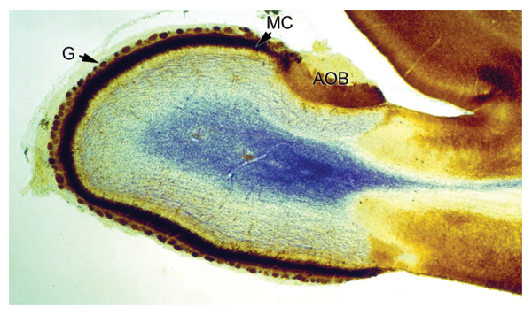
Description: Figure legend from article (CC-by): "Section through the olfactory bulb of a 16 days old rat brain. The tissue has been fixed and immunoperoxidase-stained with antibodies against GABAA-receptor_1-subunit (brown) as described elsewhere [157]. Nissl staining was performed to counter stain (blue). Clearly visible are the accessory olfactory bulb (AOB), to which chemosensory neurons from the vomeronasal organ project, the intensely labelled layer of mitral cells (MC), and the glomeruli (G), which represent the first relay station for sensory information transmitted from the nose to the brain (Jacques Paysan, unpublished)."
Author: Prepared for Commons by User:OldakQuill from a CC-by-2.0 figure in a journal article by Rebecca Elsaesser and Jacques Paysan (see source).
Usage Terms: Creative Commons Attribution-Share Alike 3.0
License: CC-BY-SA-3.0
License Link: http://creativecommons.org/licenses/by-sa/3.0/
Attribution Required?: Yes
Image usage
The following page links to this image:
