Phase-contrast microscopy facts for kids
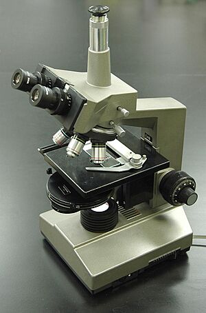
A phase-contrast microscope
|
|
| Uses | Microscopic observation of unstained biological material |
|---|---|
| Inventor | Frits Zernike |
| Manufacturer | Leica, Zeiss, Nikon, Olympus and others |
| Model | kgt |
| Related items | Differential interference contrast microscopy, Hoffman modulation-contrast microscopy, Quantitative phase-contrast microscopy |
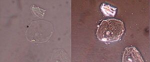
Phase-contrast microscopy (PCM) is a special way to look at tiny things using a microscope. It helps us see parts of clear objects that are usually invisible. These invisible changes in light, called "phase shifts," become visible as bright or dark areas in the image.
When light travels through something, it changes. Some changes make things brighter or darker, which we can easily see. But other changes, called phase shifts, are normally hidden. Phase-contrast microscopy helps us see these hidden changes. They give us important clues about what we are looking at.
This type of microscopy is super important in biology. It lets us see many parts inside cells that are invisible with a regular microscope. Before this invention, scientists had to stain cells to see them. But staining often killed the cells.
The phase-contrast microscope changed everything. It allowed biologists to study living cells and watch them grow and divide. It's one of the few ways to study cells without using special glowing chemicals. Because it was such a big step forward, its inventor, Frits Zernike, won the Nobel Prize in Physics in 1953.
How a Phase-Contrast Microscope Works
The main idea behind phase-contrast microscopy is clever. It separates the light that goes through the background from the light that bounces off the tiny object you are looking at. Then, it treats these two types of light differently.
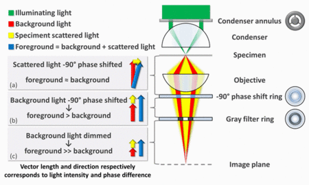
Imagine a ring of light shining through the microscope's condenser. This light then hits your specimen, like a cell. Some of this light goes straight through the specimen without changing much. This is the background light. Other light hits the specimen and gets scattered or bent. This scattered light shows the details of your specimen.
Normally, the scattered light from a clear specimen is very weak. It also has a slight "phase shift" compared to the background light. This means the foreground (your specimen) and the background look almost the same. This makes the image very hard to see.
A phase-contrast microscope fixes this in two ways. First, it makes the scattered light and background light work together. It does this by sending the background light through a special ring that shifts its phase. This makes the background light and scattered light line up perfectly.
When these two types of light meet at the image plane (where you look or where a camera is), they combine. This combining makes the areas with your specimen brighter than the areas without it. Second, the microscope also dims the background light. This makes the scattered light from your specimen stand out even more.
This process creates a clear image where the invisible parts of your specimen become visible. Most phase-contrast microscopes use "negative phase contrast," where the specimen appears brighter. In "positive phase contrast," the specimen appears darker against a lighter background.
Other Ways to See Invisible Things
The success of the phase-contrast microscope led to other cool ways to see invisible details.
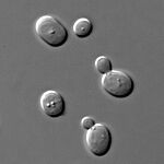
One method is called differential interference contrast (DIC) microscopy. It creates artificial shadows, making it look like the object is lit from the side. This gives a cool 3D effect. However, DIC doesn't work well if your object or its container changes how light is polarized.
Another method is Hoffman modulation contrast microscopy. This was invented in 1975 and is often used instead of DIC, especially with plastic containers.
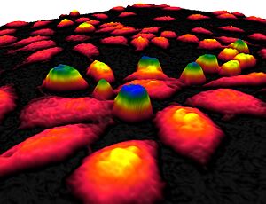
Since digital cameras became common, new digital methods have appeared. These are called quantitative phase-contrast microscopy. They create two separate images. One is a regular bright-field image. The other is a "phase-shift image." This phase-shift image actually measures the exact phase shift caused by the object. This helps scientists measure the thickness of tiny objects.
See also
- Live cell imaging
- Phase-contrast imaging
- Phase-contrast X-ray imaging
 | Emma Amos |
 | Edward Mitchell Bannister |
 | Larry D. Alexander |
 | Ernie Barnes |


