Heart valve facts for kids
Quick facts for kids Heart valve |
|
|---|---|
| System | Cardiovascular |
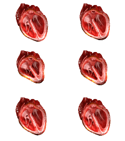
Your heart is an amazing pump! Inside your heart, there are special parts called heart valves. Think of them like one-way doors. They make sure your blood always moves in the correct direction through the different parts of your heart.
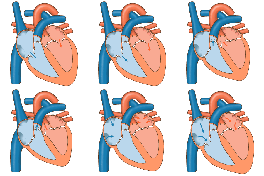
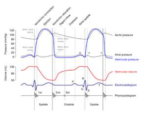
Your heart has four main valves. These valves open and close because of changes in blood pressure. When blood pushes on one side, the valve opens. When the pressure changes, it closes to stop blood from flowing backward.
The four main valves are:
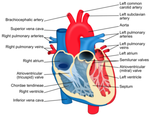
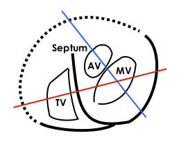
- The mitral valve and the tricuspid valve. These are between the upper chambers (called atria) and the lower chambers (called ventricles).
- The aortic valve and the pulmonary valve. These are at the exits of your heart, where blood goes into big arteries to travel to your body or lungs.
Contents
How Heart Valves Are Built
Your heart valves are amazing structures! They are covered with a smooth lining called the endocardium, which also covers the inside of your heart chambers.
Each valve has strong, flexible flaps called leaflets or cusps. These cusps open up to let blood flow through and then snap shut to create a tight seal, stopping blood from going backward.
- The mitral valve has two cusps.
- The other three valves (tricuspid, aortic, and pulmonary) each have three cusps.
We can group the heart valves into two main types:
- Atrioventricular valves: These are located between the upper and lower chambers of your heart. They stop blood from flowing back into the upper chambers (atria) when the lower chambers (ventricles) pump blood out.
- The tricuspid valve is on the right side, between the right atrium and the right ventricle.
- The mitral valve (also called the bicuspid valve) is on the left side, between the left atrium and the left ventricle.
- Semilunar valves: These are at the exits of your heart, where blood leaves the ventricles to go into large arteries. They stop blood from flowing back into the ventricles after it has been pumped out.
- The pulmonary valve is between the right ventricle and the pulmonary artery, which carries blood to your lungs.
- The aortic valve is between the left ventricle and the aorta, the body's largest artery, which carries blood to the rest of your body.
Atrioventricular Valves: The Upper-Lower Doors
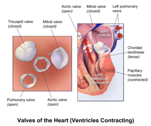
The mitral valve and the tricuspid valve are called atrioventricular valves. They sit between the upper chambers (atria) and the lower chambers (ventricles). Their job is to stop blood from flowing backward into the atria when the ventricles pump.
These valves are held in place by strong, string-like cords called chordae tendineae. Think of them like tiny parachutes attached to small muscles called papillary muscles in the heart walls. These cords and muscles work together to keep the valve flaps from flipping the wrong way when the heart squeezes.
When these atrioventricular valves close, they make the first sound of your heartbeat, which sounds like "lub".
The Mitral Valve
The mitral valve is on the left side of your heart. It has two cusps, which is why it's also called the bicuspid valve. It lets blood flow from the left atrium into the left ventricle. When the left ventricle is full and ready to pump blood to your body, the mitral valve closes tightly.
The Tricuspid Valve
The tricuspid valve is on the right side of your heart. It has three cusps. This valve is located between the right atrium and the right ventricle. It makes sure blood flows from the right atrium to the right ventricle and stops it from going backward.
Semilunar Valves: The Exit Doors
The aortic valve and the pulmonary valve are known as semilunar valves. "Semilunar" means "half-moon shaped," which describes their cusps. These valves are at the very beginning of the large arteries that carry blood away from your heart. They make sure blood flows forward into these arteries and doesn't come back into the ventricles. Unlike the atrioventricular valves, these valves do not have the string-like chordae tendineae.
When these semilunar valves close, they create the second sound of your heartbeat, which sounds like "dub".
The Aortic Valve
The aortic valve has three cusps and is located between your left ventricle and the aorta. The aorta is the main artery that sends oxygen-rich blood to your entire body. When your left ventricle pumps, the aortic valve opens to let blood rush into the aorta. Then, it quickly closes to prevent blood from flowing back into the ventricle.
The Pulmonary Valve
The pulmonary valve also has three cusps. It sits between your right ventricle and the pulmonary artery. The pulmonary artery carries blood to your lungs to pick up oxygen. When your right ventricle pumps, the pulmonary valve opens to send blood to your lungs. It then closes to stop blood from returning to the ventricle.
How Heart Valves Work
The main job of your heart valves is to control the flow of blood. They open and close at just the right time, all thanks to changes in blood pressure.
Imagine a balloon. If you squeeze one side, the air rushes to the other side. In your heart, when a chamber fills with blood, the pressure inside it goes up. This pressure pushes the valve open, letting blood move forward. Then, as the blood moves out, the pressure changes again, causing the valve to close tightly. This prevents blood from flowing backward, which would be like trying to drive a car in reverse when you want to go forward!
When Heart Valves Don't Work Right
Sometimes, heart valves don't work as perfectly as they should. This is called valvular heart disease. There are two main problems that can happen:
- Leaky Valves (Regurgitation): This happens when a valve doesn't close completely. It's like a door that doesn't shut all the way, letting some blood flow backward. This means the heart has to work harder to pump the same amount of blood.
- Narrow Valves (Stenosis): This occurs when a valve opening becomes too narrow or stiff. It's like trying to push water through a tiny straw instead of a wide pipe. The heart has to pump much harder to get blood through the narrowed opening.
These problems can affect any of the four heart valves. Sometimes, people are born with valve problems (called congenital). Other times, valve problems can develop later in life (called acquired), perhaps due to infections or other health conditions. For example, a past illness like rheumatic fever can sometimes damage heart valves.
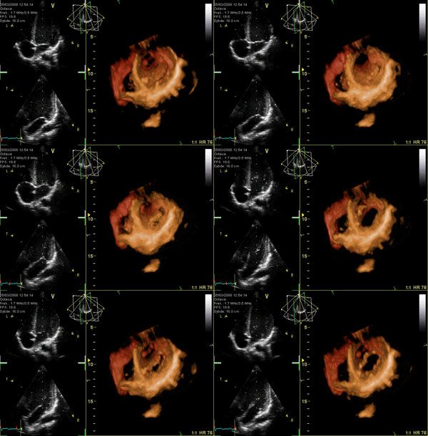
When valves don't work well, a person might feel tired, short of breath, or have swelling. Doctors can find out if there's a valve problem using a special type of ultrasound called an echocardiography. This uses sound waves to create pictures of the heart and its valves.
If a valve is badly damaged, doctors can sometimes repair it. If it's too damaged, they might need to replace it with an artificial heart valve or a valve from an animal or human donor. If an infection is causing the problem, antibiotics can help.
Valve Problems from Birth
Sometimes, a baby is born with a heart valve that isn't formed correctly. This is called a congenital heart defect.
One common example is a bicuspid aortic valve. Remember, the aortic valve usually has three cusps. But in this condition, two of the cusps are fused together, making it a two-cusp valve instead of three. This can sometimes cause the valve to become narrow over time.
Other times, a valve might be completely missing, or its parts might be in the wrong place. Doctors can often find these problems early and help treat them.
A Look Back: The History of Heart Valves
People have been curious about the heart for a very long time! Over 500 years ago, the famous artist and inventor Leonardo da Vinci was one of the first to carefully study and draw heart valves. He learned about them by looking at animal and human hearts. He even made models out of wax and glass to understand how blood flowed through them.
Artificial Heart Valves
For many years, if someone had a damaged heart valve, there wasn't much doctors could do. But in 1960, a big step forward happened! The first successful artificial heart valve was invented by Miles "Lowell" Edwards. It was called the Star-Edwards valve. This amazing invention helped many people whose own heart valves were no longer working.
Since then, doctors and scientists have continued to create even better artificial valves. These new valves have helped countless people live healthier lives.
See also
 In Spanish: Válvula cardiaca para niños
 | James B. Knighten |
 | Azellia White |
 | Willa Brown |


