
Ascaris lumbricoides facts for kids
Quick facts for kids Ascaris lumbricoides |
|
|---|---|
 |
|
| An adult female Ascaris worm | |
| Scientific classification |
|
| Kingdom: | Animalia |
| Phylum: | Nematoda |
| Class: | Chromadorea |
| Order: | Ascaridida |
| Family: | Ascarididae |
| Genus: | Ascaris |
| Species: |
A. lumbricoides
|
| Binomial name | |
| Ascaris lumbricoides Linnaeus, 1758
|
|
| Script error: The function "autoWithCaption" does not exist. | |
Script error: No such module "Check for conflicting parameters".
Ascaris lumbricoides is a type of roundworm that lives in humans. It's often called the "large human roundworm" because it can grow quite long, up to 35 centimeters (about 14 inches). This makes it one of the biggest worms that can live inside people.
It belongs to a group of worms called nematodes. Ascaris lumbricoides is the most common parasitic worm found in humans worldwide. It causes a disease called ascariasis. This disease is especially common in warm, tropical, and subtropical countries. Scientists think that about one-sixth of all people in the world might have this worm.
Some scientists believe that Ascaris lumbricoides and Ascaris suum, which is a roundworm found in pigs, might actually be the same species.
Contents
How the Worm Lives and Spreads
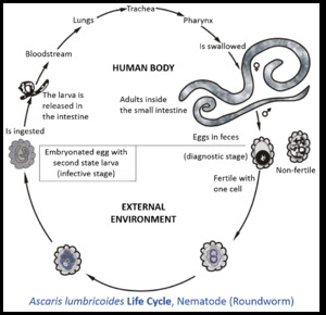
Ascaris lumbricoides spreads from person to person when tiny eggs from the worm are swallowed. This often happens through contaminated food or water.
Here's how its life cycle works:
- Adult female worms live in a person's intestines and lay many eggs.
- These eggs leave the body in a person's poop.
- If the eggs are fertilized, they need to spend some time in warm, moist soil to become infective. This can take a few weeks.
- When someone accidentally swallows these infective eggs (for example, if they eat food grown in contaminated soil or don't wash their hands), the eggs hatch inside their small intestine.
- Tiny worm larvae then break through the intestine wall and enter the bloodstream.
- They travel through the blood to the liver and heart, then move into the lungs.
- In the lungs, they grow a bit more and then move up the breathing tubes to the throat.
- From the throat, they are swallowed again, returning to the small intestine.
- Once back in the small intestine, they grow into adult worms.
- Female worms can then start laying up to 200,000 eggs every day for over a year!
- These eggs are very tough. They have a special outer layer that protects them from harsh chemicals like acids. This helps them survive in the soil for a long time, sometimes even for 10 years or more.
What the Worm Looks Like


Ascaris lumbricoides worms are quite large.
- Male worms are usually about 15 to 31 centimeters (6 to 12 inches) long and about 2 to 4 millimeters (0.08 to 0.16 inches) thick. Their tail end is curved.
- Female worms are even bigger, growing to about 20 to 49 centimeters (8 to 19 inches) long and 3 to 6 millimeters (0.12 to 0.24 inches) thick. They can hold millions of eggs inside them at one time.
The eggs are also distinct:
- Fertilized eggs are oval or round. They are very tiny, about 45 to 75 micrometers long (you need a microscope to see them). They have a thick outer shell.
- Unfertilized eggs are a bit different in shape and size.
Where Infections Are Common
Many people around the world are infected with Ascaris lumbricoides. It's most common in parts of Africa, the Americas, China, and East Asia. Even though it's less common in places like the United States, it can still be found in warmer, humid areas.
The eggs of Ascaris lumbricoides are very strong. They can survive in soil for many months or even years, even in very cold or dry conditions. Scientists have found these ancient worm eggs in old human waste samples from over 24,000 years ago, showing how long this parasite has been around.
About the Infection (Ascariasis)
Infections with these worms are more common in places where sanitation is poor. This means areas where there aren't good systems for treating human waste. Sometimes, raw human waste is used as fertilizer for crops, which can spread the eggs.
Symptoms of Infection
Often, if someone has only a few worms, they might not feel sick at all. Sometimes, the only sign is seeing a worm pass out of the body.
If someone has a lot of worms, they might experience symptoms like:
- Coughing
- Fever
- Tummy pain or discomfort
- Sometimes, passing the long worms
In some cases, the worms can cause more serious problems, like blockages in the intestines. When the tiny worms travel through the lungs, they can sometimes cause lung problems or allergic reactions.
How to Prevent Infection
Preventing Ascaris infection is mostly about good hygiene and proper waste management.
- Wash hands: Always wash your hands thoroughly with soap and water, especially before eating or preparing food, and after using the bathroom.
- Clean food: Wash fruits and vegetables carefully, especially if they might have been grown in soil fertilized with human waste. Cooking food thoroughly also kills the eggs.
- Safe waste disposal: Proper sewage systems and treatment of human waste are very important to stop the spread of eggs.
- Egg resistance: Ascaris eggs are very hard to kill. They can survive many common cleaning products like bleach. High heat, like cooking or very hot composting (over 50°C or 122°F for 24 hours), can kill them.
Details of How Infection Happens
When a person swallows the tiny, infective eggs, they hatch in the small intestine. The young worms then travel through the body:
- They first enter the bloodstream.
- They pass through the right side of the heart and into the lungs.
- In the lungs, they break out of tiny blood vessels and enter the air spaces.
- From the lungs, they crawl up the breathing tubes to the throat.
- They are then swallowed again, returning to the small intestine.
- Once in the small intestine, they grow into adult worms and start laying eggs within about two months.
It might seem strange that the worms travel all around the body just to end up where they started. One idea is that this journey helps them grow bigger and stronger, which allows them to lay more eggs.
History of Discovery
People have known about these giant intestinal worms for a very long time. In 1758, a famous scientist named Carl Linnaeus gave them their scientific name, Ascaris lumbricoides.
For many centuries, people didn't understand how these worms appeared. They thought the worms just grew on their own inside the body. But in the mid-1800s, scientists started to figure it out.
- In 1855, Henry Ransom in England found Ascaris eggs in human poop.
- Later, in 1886, Salvatore Calandruccio in Italy successfully infected a boy by giving him Ascaris eggs, showing how the infection starts.
- Over the next few decades, more scientists, like Francis Stewart and Sadao Yoshida, did important research. They studied how the worms traveled through the body in animals and even in themselves!
- Finally, in 1922, Shimesu Koino bravely swallowed 2,000 Ascaris lumbricoides eggs. He found the young worms in his spit a few days later, and then adult worms in his poop after taking medicine. This experiment fully confirmed the worm's life cycle.
Images for kids
-
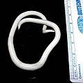
An adult female Ascaris worm
-
Image showing lifecycle inside and outside of the human body of one fairly well described helminth: A. lumbricoides
-
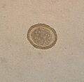
Fertile egg as can be seen in a microscope
-
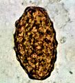
Infertile egg
See also
 In Spanish: Ascaris lumbricoides para niños
