Christine P. Hendon facts for kids
Quick facts for kids
Christine P. Hendon
|
|
|---|---|
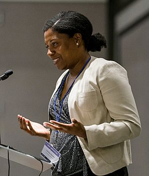
Hendon in 2019
|
|
| Nationality | American |
| Alma mater | Massachusetts Institute of Technology Case Western Reserve University Harvard Medical School |
| Known for | Optical coherence imaging for interventional heart arrhythmia procedures |
| Awards | 2017 Presidential Early Career Awards for Scientists and Engineers (PECASE), 2013 MIT Technology Review’s 35 Innovators Under 35, 2012 Forbes 30 under 30 in Science and Healthcare |
| Scientific career | |
| Fields | Electrical and biomedical engineering |
| Institutions | Columbia University |
Christine P. Hendon is an amazing engineer and computer scientist. She is a professor at Columbia University in New York City. Dr. Hendon is a leader in medical imaging. She creates special tools that use light to see inside the body. These tools help doctors guide treatments and understand how body parts work. Her new imaging methods help doctors find and treat heart problems, breast cancer, and even understand why some babies are born too early. She has won many awards, including being named one of Forbes' "30 Under 30" and receiving a special award from President Barack Obama in 2017.
Contents
Early Life and School
Christine Hendon, whose maiden name was Fleming, wanted to be a teacher when she was a kid. In high school, she loved math and science. She joined a program with NASA that studied climate and planets. This program made her want to become a scientist.
In 2000, Dr. Hendon went to college at the Massachusetts Institute of Technology (MIT) in Cambridge, Massachusetts. She studied Electrical Engineering and Computer Science. She started doing research right away. She earned her bachelor's degree in 2004.
After MIT, Dr. Hendon went to Case Western Reserve University. There, she earned her Master's degree in 2007 and her PhD in Biomedical Engineering in 2010. For her PhD, she worked on a special imaging method called Optical coherence tomography (OCT). She used OCT to create 3D pictures of human tissues and organs. This helped doctors treat heart problems called cardiac arrhythmias. She even created a computer program to help understand changes in heart tissue. Her work showed that OCT could help doctors see what they were doing during heart treatments, making them more successful.
After her PhD, Dr. Hendon did more research at Harvard Medical School and Massachusetts General Hospital. She worked on making OCT even better. She finished this research in 2012.
Career and Research
In 2012, Dr. Hendon became a professor at Columbia University. In 2018, she became an Associate Professor. She leads a lab called the Structure-Function Imaging Laboratory. Her lab creates new medical tools that use imaging to help doctors diagnose and treat cancer and heart problems. Her work uses computer programs to get important information from OCT images in real-time. Dr. Hendon is also a member of several important science and engineering groups.
Helping Heart Treatments with OCT
Dr. Hendon helped improve how doctors treat a heart problem called atrial fibrillation. She used a special catheter (a thin tube) with a light-based imaging system. Her work showed that this helped make heart treatments more successful.
She also used her OCT skills to understand how heart tissue is built and how it works. She could see tiny details like elastic fibers and collagen. Since the way heart tissue is made affects how diseases start and how people recover, Dr. Hendon and her team created a computer method to figure out what heart tissue is made of using OCT. This method was over 80% accurate. Thanks to her technology, doctors can now see changes in heart tissue in real-time during treatment. This helps them be more accurate and improves how patients recover.
OCT for Breast Cancer
Dr. Hendon also started using her OCT methods to help diagnose and treat breast cancer. This imaging technique is sometimes called "optical ultrasound." Using very high-resolution OCT, she made it easier to find and understand breast cancer.
OCT Imaging and Preterm Birth
Dr. Hendon then became interested in understanding the cervix, which is part of the female reproductive system. She wanted to see how its structure affects why some babies are born too early (preterm birth). She found that the way collagen fibers are arranged in the cervix affects how it changes during pregnancy. Since these changes can lead to the cervix shortening, which might cause preterm birth, Dr. Hendon wanted to understand the tiny structural details that cause this shortening. By looking at the collagen fibers with OCT, she could figure out why the cervix changes shape. This helps scientists understand the causes of preterm birth at a very tiny level.
Awards and Honors
- 2021 Elected as Fellow of the International Society for Optics and Photonics (SPIE)
- 2017 Presidential Early Career Awards for Scientists and Engineers (PECASE)
- 2015 National Science Foundation Career Award
- 2015 Rodriguez Family Junior Faculty Development Award at the School of Engineering and Applied Science
- 2014 NIH New Innovator Award
- 2013 MIT Technology Review's 35 Innovators Under 35
- 2012 Forbes 30 under 30 in Science and Healthcare
- 2012 Wellman-Bullock Postdoctoral Fellowship - Massachusetts General Hospital
 | Georgia Louise Harris Brown |
 | Julian Abele |
 | Norma Merrick Sklarek |
 | William Sidney Pittman |

